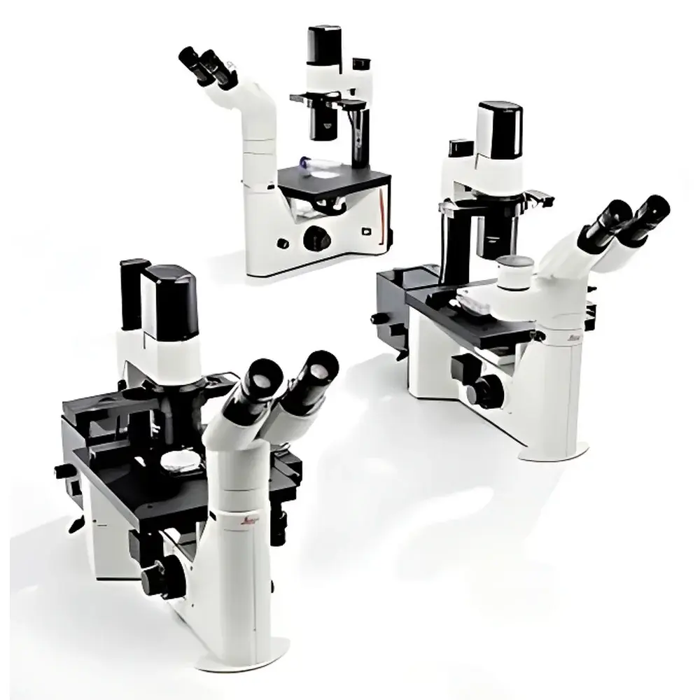
Leica DM IL LED Inverted Microscope
| Brand | Leica |
|---|---|
| Origin | Germany |
| Model | Leica DM IL LED |
| Illumination | 10 W high-intensity LED |
| LED Lifetime | 50,000 hours |
| Contrast Method | Integrated adjustable Phase Contrast (Ph1–Ph4 compatible) |
| Working Distance | Extended for large culture vessels (e.g., T-flasks, Petri dishes, multi-well plates) |
| Stage | Height-adjustable mechanical stage with X–Y translation |
| Eyepiece Tube | Ergonomic binocular tube with adjustable interpupillary distance and diopter correction |
| Compliance | CE-marked, compliant with IEC 61000-6-3 (EMC) and IEC 61000-6-2 (immunity), designed for ISO 13485-aligned laboratory environments |
| Accessories Support | Compatible with optional heated stage, fluorescence LED modules (e.g., GFP, RFP, DAPI excitation), fiber-optic illuminators, and micromanipulation systems |
Overview
The Leica DM IL LED is a robust, entry-level inverted microscope engineered specifically for routine live-cell observation, tissue culture monitoring, and basic micro-manipulation workflows in academic, clinical, and industrial life science laboratories. Built on Leica’s legacy of precision optical engineering, the system employs an optimized Köhler illumination pathway coupled with a high-efficiency 10 W white LED light source—delivering stable color temperature (~5,700 K), minimal thermal load on specimens, and consistent photometric output across its 50,000-hour operational lifetime. Unlike halogen-based systems, the LED illumination eliminates the need for warm-up time, filter wheel synchronization, or frequent bulb replacement, thereby reducing maintenance overhead and improving experimental reproducibility. The inverted configuration places the objective lenses beneath the specimen stage—enabling direct access to cell cultures housed in standard flasks, Petri dishes, or multi-well plates without disturbing sterility or requiring sample inversion. Its optical path integrates fully corrected achromatic objectives (typically 10×, 20×, 32×, and 40× Ph) and a phase contrast slider mechanism that supports Ph1 through Ph4 settings—allowing rapid, repeatable contrast optimization without dedicated phase objectives.
Key Features
- Ergonomic design with height-adjustable stage and binocular observation tube—supports customizable interpupillary distance, diopter compensation, and viewing angle for prolonged user comfort.
- Integrated, motor-free phase contrast adjustment lever—enables intuitive, tool-free switching between phase rings during live imaging sessions.
- Extended working distance (≥50 mm at 10×, ≥25 mm at 40×)—accommodates tall culture vessels, perfusion chambers, and environmental control enclosures without mechanical interference.
- LED illumination with analog/digital brightness control—provides linear intensity regulation from 10% to 100%, synchronized automatically with selected phase contrast setting to maintain optimal signal-to-noise ratio.
- Modular platform architecture—accepts third-party and Leica-certified accessories including temperature-controlled stages (37°C ±0.5°C), LED fluorescence excitation modules (GFP/DAPI/RFP), and fiber-optic side-illumination ports for transmitted-light enhancement.
- Compliance-ready mechanical construction—designed for integration into GLP- and GMP-aligned workflows; documentation supports traceability per ISO/IEC 17025 requirements when used with calibrated reference standards.
Sample Compatibility & Compliance
The Leica DM IL LED is validated for use with adherent and suspension mammalian cell lines (e.g., HeLa, CHO-K1, NIH/3T3), primary neurons, zebrafish embryos, and 3D organoid cultures grown in standard polystyrene or glass-bottom vessels. Its open-stage geometry permits simultaneous integration of micropipette holders, patch-clamp rigs, or microinjection systems—facilitating electrophysiology and CRISPR delivery protocols. From a regulatory standpoint, the instrument conforms to EN 61000-6-2 (electromagnetic immunity) and EN 61000-6-3 (emission limits), carries CE marking under Directive 2014/30/EU (EMC) and 2014/35/EU (LVD), and meets essential safety requirements outlined in IEC 61010-1 for laboratory equipment. While not FDA 510(k)-cleared as a diagnostic device, it satisfies foundational performance criteria referenced in CLSI EP17-A2 for optical microscopy validation in QC/QA contexts.
Software & Data Management
The DM IL LED operates as a hardware platform compatible with Leica Application Suite (LAS) X Core software (sold separately), enabling image capture, annotation, measurement (area, length, intensity profiling), and time-lapse acquisition with metadata tagging (user ID, timestamp, objective, illumination mode). When paired with LAS X, the system supports audit-trail generation compliant with FDA 21 CFR Part 11 requirements—including electronic signatures, session logs, and immutable data export (TIFF, JPEG2000, or Leica’s native .lei format). Raw image files retain embedded EXIF-like metadata: exposure time, gain, LED intensity percentage, and stage coordinates—ensuring full traceability for ISO 13485 internal audits or external accreditation reviews.
Applications
- Routine quality control of cell monolayers in biomanufacturing (e.g., passage verification, confluency assessment, contamination screening).
- Long-term time-lapse imaging of proliferation, migration, and morphological dynamics in primary and stem cell cultures.
- Support for manual microinjection, nuclear transfer, and oocyte manipulation in reproductive biology labs.
- Pre-screening of fluorescent reporter expression prior to flow cytometry or sequencing workflows.
- Teaching laboratories—where reliability, ease of alignment, and low total cost of ownership support high-throughput student training in basic microscopy principles.
FAQ
Does the Leica DM IL LED support fluorescence imaging?
Yes—via optional LED fluorescence modules (e.g., Leica LUM020 for GFP, LUM030 for RFP) or fiber-optic coupling to external mercury/xenon sources. No built-in fluorescence capability is included in the base configuration.
Is the LED illumination intensity adjustable during live imaging?
Yes—intensity is continuously variable via front-panel rotary encoder or software command, with real-time feedback displayed on the LED driver status indicator.
Can the microscope be integrated into a cleanroom environment?
Yes—the unit is constructed with non-outgassing materials, has no internal cooling fans, and complies with ISO 14644-1 Class 7 particulate limits when operated within laminar flow hoods or isolators.
What is the maximum recommended magnification for quantitative phase contrast measurements?
For optimal resolution and depth-of-field balance, 40× Ph objectives are routinely used; higher magnifications (e.g., 63×) require oil immersion and are not supported by the standard DM IL LED optical train.
Are calibration certificates available for metrological traceability?
Leica offers optional NIST-traceable stage micrometer and eyepiece graticule calibration kits (order codes: 11506295, 11506296), accompanied by ISO/IEC 17025-accredited calibration reports upon request.
Related Products


