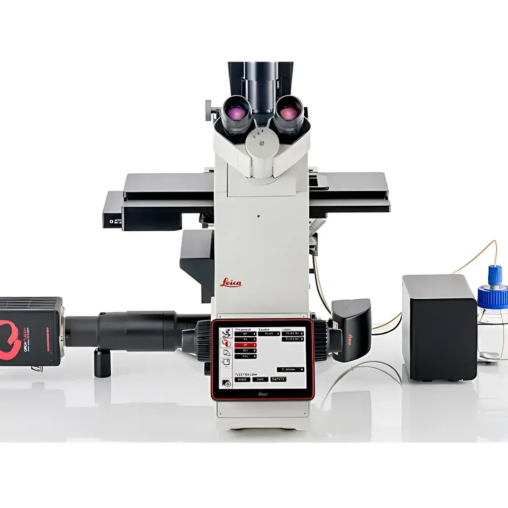
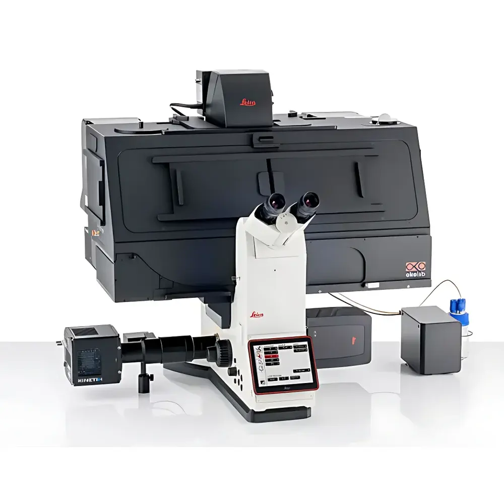
Leica DMi8 Inverted Microscope
| Brand | Leica |
|---|---|
| Origin | Shanghai, China |
| Manufacturer Type | Authorized Distributor |
| Product Origin | Domestic (China) |
| Model | DMi8 |
| Pricing | Upon Request |
Overview
The Leica DMi8 is a high-performance inverted research microscope engineered for demanding live-cell imaging, 3D tissue analysis, and quantitative microscopic image analysis. Built on a modular optical platform, it integrates advanced illumination, precision mechanics, and computational imaging technologies—including THUNDER™ Computational Clearing, Adaptive Focus Control (AFC), and SmartCORR™—to deliver high-fidelity, reproducible data from thick, dynamic, and physiologically relevant specimens. Its optical design adheres to Köhler illumination principles and supports both widefield and structured illumination modalities. The system is optimized for applications requiring long-term stability under controlled environmental conditions (e.g., CO₂, temperature, humidity), making it suitable for GLP-compliant cell culture monitoring, developmental biology, neuroimaging, and preclinical tissue phenotyping.
Key Features
- 22 mm Field of View (FOV) Optics: Enables rapid acquisition of large-area overviews with high spatial uniformity—critical for multi-position screening across slides, dishes, and multiwell plates.
- Adaptive Water Immersion Technology: Patented sensor-driven immersion maintenance ensures consistent refractive index matching during extended time-lapse experiments, enabling reliable use of water-dipping objectives (e.g., 40×/1.10 W, 63×/1.20 W) without manual rehydration or sample displacement.
- Integrated Sample Finder: Automates whole-sample overview generation in seconds; captures focus-stacked mosaics directly from the stage, eliminating manual search and enabling precise spatial registration of acquired datasets.
- Enhanced Navigator Workflow: Combines template-based experiment definition (for standard vessel formats) with seamless transition from low-magnification overviews to high-resolution immersion objectives—without moving the sample off-stage.
- THUNDER Imaging Platform: Real-time Computational Clearing algorithm removes out-of-focus blur from widefield and spinning-disk confocal acquisitions, delivering optical-sectioning quality without the phototoxicity or acquisition time penalties of point-scanning systems.
- Quantum Precision Stage & Hardware Synchronization: Fully motorized XYZ stage with sub-micron repeatability; supports TTL-triggered acquisition synchronization with cameras, light sources, and environmental controllers for robust multimodal workflows.
Sample Compatibility & Compliance
The DMi8 accommodates standard and custom specimen carriers including glass-bottom dishes (35 mm, 60 mm), chambered coverslips, multiwell plates (6–384-well), and histological slides. It supports live-cell assays under physiological conditions via optional incubation chambers compliant with ISO 13485–certified environmental control modules. All imaging workflows can be configured to meet audit-trail requirements per FDA 21 CFR Part 11 when paired with Leica Application Suite X (LAS X) software in validated mode. System documentation conforms to ISO/IEC 17025 guidelines for instrument qualification and routine performance verification.
Software & Data Management
Leica LAS X software provides unified control of hardware, acquisition, analysis, and metadata annotation. It includes built-in support for FAIR data principles—automated embedding of MIAME/MINSEQE-compliant metadata, DICOM-SR export for pathology integration, and HDF5-based storage for scalable large-volume dataset handling. The software supports GLP/GMP audit trails with user-level permissions, electronic signatures, and immutable logging of all acquisition parameters and processing steps. THUNDER Live enables real-time parameter optimization with immediate visual feedback, reducing experimental iteration cycles. LAS X also interfaces with third-party analysis platforms (e.g., Imaris, Fiji, Python-based pipelines) via open APIs and standardized file formats (OME-TIFF, ND2, CZI).
Applications
- Long-term live-cell imaging of organoids, spheroids, and co-cultures under controlled gas and thermal environments.
- High-content screening of drug responses in adherent and suspension cell models using multi-channel fluorescence and phase contrast.
- 3D reconstruction and quantitative morphometry of cleared tissue sections (e.g., mouse brain, lung, kidney) up to 200 µm thickness.
- Subcellular dynamics studies using TIRF and spinning-disk confocal modules—particularly for membrane trafficking, cytoskeletal remodeling, and nuclear envelope dynamics.
- Correlative light-electron microscopy (CLEM) preparation workflows leveraging precise coordinate mapping between overview and high-mag fields.
- Teaching and core facility deployment due to intuitive workflow templates, remote operation capability, and role-based access controls.
FAQ
Is the DMi8 compatible with third-party cameras and detectors?
Yes—the system features standard C-mount and F-mount interfaces, along with GenICam-compliant driver support for major scientific CMOS and EMCCD cameras.
Can THUNDER processing be applied retrospectively to existing widefield datasets?
Yes—LAS X allows offline THUNDER deconvolution of archived OME-TIFF or LSM files, provided raw z-stack data and objective metadata are preserved.
What environmental control options are available for long-term live imaging?
Leica offers integrated CO₂/O₂/temperature/humidity modules certified for ISO Class 5 cleanroom compatibility, with PID-controlled regulation and real-time sensor logging.
Does the DMi8 support automated focus drift correction during time-lapse?
Yes—Adaptive Focus Control (AFC) uses infrared reflection detection to maintain focus position within ±50 nm over multi-hour acquisitions.
How is system validation handled for regulated laboratories?
Leica provides IQ/OQ documentation packages aligned with ASTM E2500 and USP , including calibration certificates traceable to NIST standards and installation verification checklists.


