NARISHIGE CP-1 Stainless Steel Chamber Plate for Chronic Stereotaxic Head Fixation in Mice
| Brand | NARISHIGE |
|---|---|
| Origin | Japan |
| Model | CP-1 |
| Material | Stainless Steel |
| Surface Treatment | Anodized Aluminum with Electrical Insulation |
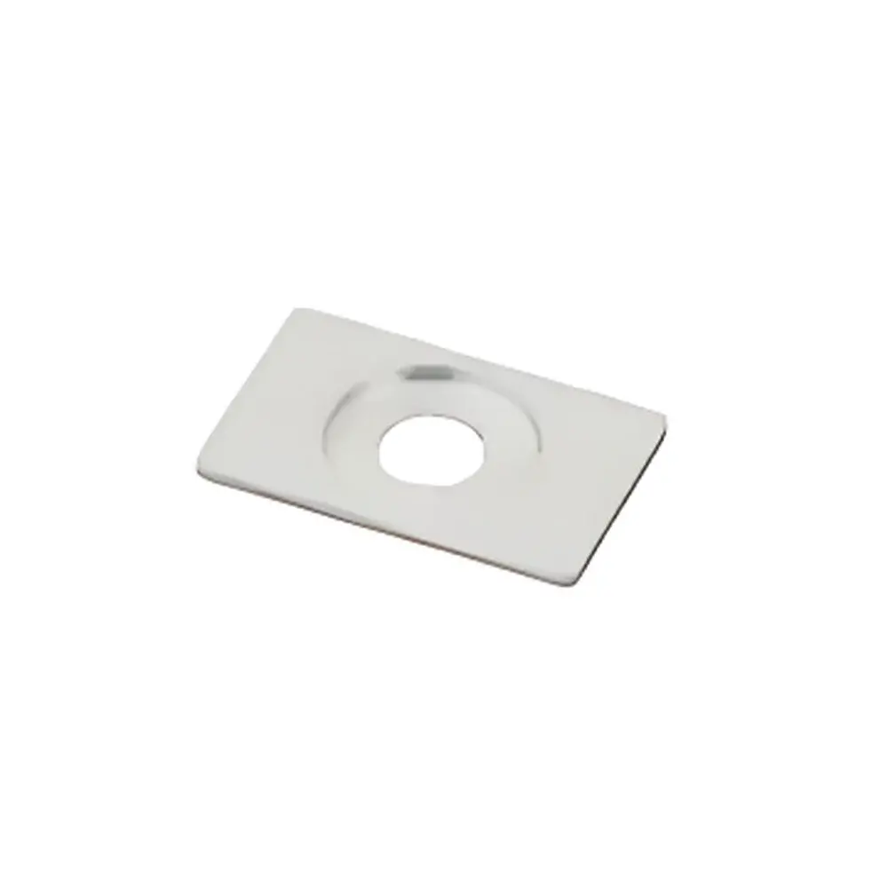
| Observation Hole Diameter | 5 mm |
| Countersunk Mounting Hole Diameter | 10 mm |
| Dimensions (W × D × T) | 12 mm × 19 mm × 1 mm |
| Unit Weight | ~0.5 g |
| Package Quantity | 5 pcs per pack |
Overview
The NARISHIGE CP-1 Stainless Steel Chamber Plate is a precision-engineered component designed for chronic stereotaxic head fixation in murine neuroscience research. It functions as a mounting interface between the animal’s skull and external recording or stimulation hardware—such as microdrives, optrodes, or electrophysiology connectors—enabling stable, repeatable positioning across longitudinal experimental sessions. Constructed from medical-grade stainless steel and finished with anodized aluminum insulation, the CP-1 provides both mechanical robustness and electrical isolation critical for low-noise electrophysiological recordings and artifact-free optical imaging. Its compact footprint (12 mm × 19 mm × 1 mm) and ultra-lightweight design (~0.5 g) minimize biomechanical load on the cranial implant site, supporting long-term viability and behavioral integrity in awake, freely moving or head-fixed paradigms. The plate operates within the broader NARISHIGE stereotaxic ecosystem—including compatible frames (e.g., SR-1, MO-10), manipulators (e.g., MO-10, MM-10), and chronic cannula systems—and is intended for use following standard craniotomy and skull-screw anchoring protocols.
Key Features
- Stainless steel substrate ensures high tensile strength, corrosion resistance, and biocompatibility under chronic in vivo conditions.
- Anodized aluminum surface coating provides uniform electrical insulation (dielectric strength ≥ 100 V/µm), reducing capacitive coupling and signal interference during extracellular or local field potential recordings.
- Integrated 5 mm diameter observation port enables real-time visual verification of dura condition, electrode tip placement, or fiber optic alignment without disassembly.
- 10 mm countersunk mounting hole accommodates standard M2.5 or #0-80 screws used in NARISHIGE cranial fixtures, ensuring flush integration with the skull surface and minimizing soft-tissue irritation.
- Dimensionally optimized geometry (12 × 19 × 1 mm) balances structural rigidity with minimal cranial coverage, preserving access to adjacent cortical or subcortical targets.
- Supplied sterile-ready; compatible with autoclaving (up to 134°C, 3 bar) and ethanol/isopropanol disinfection protocols per ISO 17664 guidelines.
Sample Compatibility & Compliance
The CP-1 is validated for use in C57BL/6, BALB/c, and CD-1 mice aged ≥8 weeks with fully ossified calvaria. It complies with ASTM F2129–22 (Standard Test Method for Conducting Cyclic Potentiodynamic Polarization Measurements to Determine the Corrosion Susceptibility of Small Metallic Implants) for metallic implant safety assessment. While not a standalone medical device, its material composition and surface treatment meet ISO 10993-5 (cytotoxicity) and ISO 10993-10 (irritation/sensitization) requirements when implanted according to established surgical best practices. The design supports GLP-compliant chronic studies requiring audit-trail documentation of implant lot numbers, sterilization cycles, and intraoperative positioning metadata.
Software & Data Management
As a passive mechanical interface, the CP-1 does not incorporate embedded electronics or firmware. However, it integrates seamlessly into digital experimental workflows via compatibility with NARISHIGE’s optional digital stereotaxic planning tools (e.g., STP-1 software export modules) and third-party platforms including OpenEphys, SpikeGLX, and Bonsai. Positional metadata—such as AP/ML coordinates relative to bregma, implant angle, and screw torque values—can be logged directly into electronic lab notebooks (ELNs) compliant with 21 CFR Part 11 when paired with timestamped surgical documentation. Traceability is enhanced by batch-specific packaging labels containing manufacturer lot codes and RoHS/REACH compliance indicators.
Applications
- Chronic electrophysiology: Stable anchoring point for silicon probes, tetrodes, or Neuropixels arrays in multi-week recording experiments.
- Optogenetics: Rigid platform for chronic fiber-optic cannulae (e.g., Doric Lenses, Thorlabs CFM series) enabling repeated light delivery with sub-50 µm positional repeatability.
- Two-photon imaging: Minimizes motion artifacts during awake head-fixed calcium imaging by eliminating micromotion at the skull-implant interface.
- Behavioral neuropharmacology: Facilitates repeated intracranial microinjections through chronically implanted guide cannulae secured via CP-1-mounted retention rings.
- Developmental neuroscience: Enables longitudinal tracking of circuit maturation in juvenile mice due to low-profile design and minimal inflammatory response.
FAQ
Is the CP-1 compatible with non-NARISHIGE stereotaxic frames?
Yes—its 10 mm countersunk hole and planar geometry allow adaptation to Leica, Stoelting, and Kopf frames using custom spacers or threaded adapters; however, optimal alignment requires verification with frame-specific coordinate mapping.
Can the CP-1 be reused after explantation?
No—per ISO 14971 risk management principles, it is designated for single-animal, single-study use to prevent cross-contamination and mechanical fatigue-induced failure.
Does the anodized layer affect MRI compatibility?
The aluminum oxide layer is non-ferromagnetic and introduces negligible susceptibility artifacts; the stainless steel base remains MRI-safe at ≤7T when fully secured and free of loose particulates.
What adhesive is recommended for skull bonding?
Dental acrylic (e.g., C&B Metabond, Ortho-Jet) applied with controlled pressure and curing under nitrogen atmosphere yields optimal shear strength (>15 MPa) and thermal stability over 4+ weeks.
How should the observation port be cleaned between procedures?
Sterile saline irrigation followed by lint-free wipe with 70% isopropanol; avoid ultrasonic cleaning or abrasive solvents that may degrade the anodized dielectric layer.


