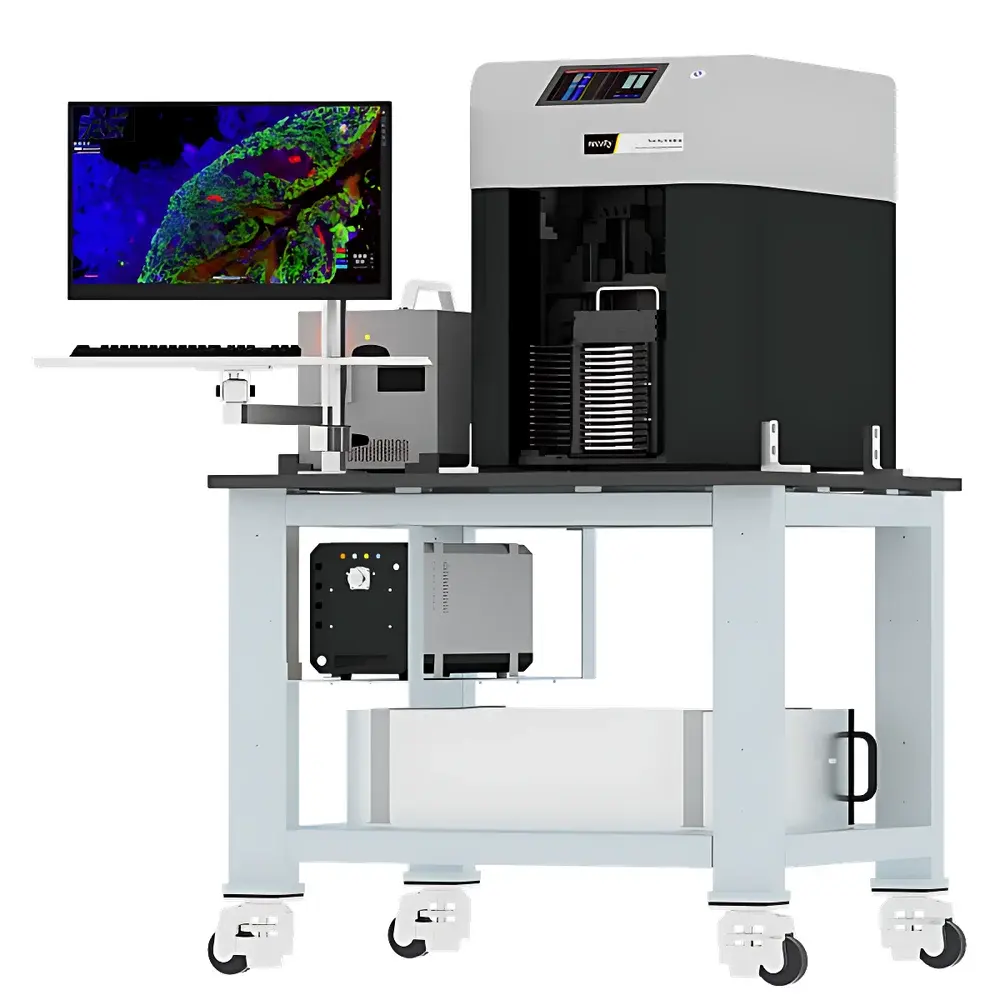
Revvity Hawkeye High-Speed, High-Resolution Whole-Slide Microscopy Scanner
| Brand | Revvity |
|---|---|
| Origin | Jiangsu, China |
| Manufacturer Type | Original Equipment Manufacturer (OEM) |
| Regional Classification | Domestic (PRC) |
| Model | Hawkeye |
| Pricing | Available Upon Request |
Overview
The Revvity Hawkeye is a high-speed, high-resolution whole-slide microscopy scanner engineered for quantitative digital pathology and advanced life science research. It employs a precision motorized stage combined with a high-NA (1.42) oil-immersion objective and sCMOS imaging architecture to deliver true subcellular spatial resolution across full tissue sections. Unlike conventional slide scanners that rely on sequential low-magnification survey scans followed by targeted high-magnification acquisition, the Hawkeye acquires native 100× oil-immersion data across the entire slide in a single pass—eliminating registration artifacts and manual intervention. Its optical design adheres to diffraction-limited performance principles, enabling reliable visualization of nuclear morphology, chromatin texture, membrane protrusions, and organelle-level features without interpolation or upscaling. The system is built around a closed-loop feedback-controlled illumination and focus architecture, ensuring consistent signal-to-noise ratio and Z-stack reproducibility across multi-day acquisition campaigns.
Key Features
- Full-slide 100× oil-immersion scanning completed in ≤180 seconds—60× faster than legacy widefield slide scanners
- Optical resolution limited only by diffraction: XY resolution < 200 nm at 100×/1.42 NA, validated per ISO 19003:2021 standards for microscope resolution testing
- Proprietary CSFA (Continuous Smart Focus Algorithm) autofocus engine delivering sub-micron Z-axis repeatability (< ±0.15 µm) across heterogeneous tissue topographies
- Real-time AI-accelerated analysis pipeline processing raw image streams at sustained rates of 10–100 Gbps via FPGA-assisted preprocessing and GPU-optimized inference kernels
- Automated classification of ≥95 morphologically distinct cell phenotypes—including rare circulating tumor cells, activated microglia, and mitotic figures—using transfer-learned convolutional neural networks trained on multi-institutional histopathology datasets
- Native support for single-cell spatial indexing: each segmented object retains full coordinate metadata, enabling retrospective re-extraction of original pixel data, intensity profiles, and spectral signatures
Sample Compatibility & Compliance
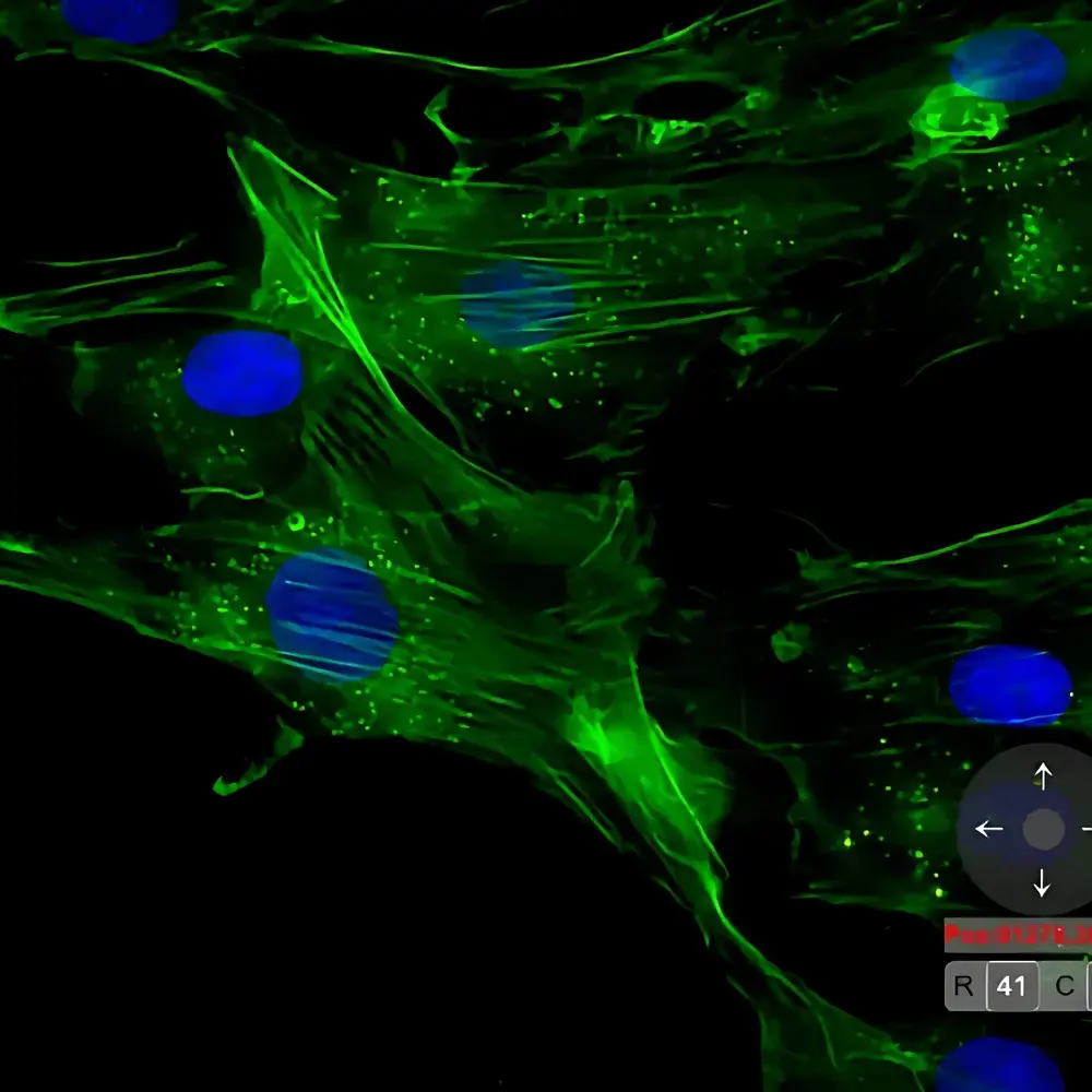
The Hawkeye accommodates standard 1×3 inch glass slides (75 × 25 mm), including those prepared with H&E, IHC, IF, multiplex immunofluorescence (up to 7-plex), and CISH protocols. It supports both dewaxed and non-dewaxed sections, as well as live-cell chamber slides (with environmental control integration). All hardware components comply with IEC 61000-6-3 (EMC emissions) and IEC 61000-6-2 (immunity) standards. The software platform meets FDA 21 CFR Part 11 requirements for electronic records and signatures, including audit trail logging, role-based access control, and immutable data archiving. Validation documentation supports GLP and GCP compliance for preclinical imaging studies.
Software & Data Management
Hawkeye Control Suite v4.x provides integrated acquisition, reconstruction, annotation, and analytics workflows. Raw data are stored in OME-TIFF format compliant with Bio-Formats 6.10+ specifications. The system includes DICOM-SR export capability for PACS integration and supports MIAME-compliant metadata embedding. Analysis modules generate FAIR-compliant outputs—including CSV tables with morphometric descriptors (area, eccentricity, solidity, Haralick texture), interactive UMAP/t-SNE projections, and flow cytometry–style scatter plots with gating overlays. All processing pipelines are containerized (Docker) and version-controlled via GitLab CI/CD, enabling reproducible computational experiments across distributed labs.
Applications
- Quantitative digital pathology: tumor grading, lymphocyte infiltration scoring (TILs), and spatial heterogeneity mapping in FFPE biopsies
- Neuroscience: high-throughput quantification of dendritic spine density, amyloid plaque burden, and microglial activation states in rodent brain sections
- Drug discovery: longitudinal monitoring of cellular phenotypic responses in 3D organoid cultures under compound treatment
- Clinical trial biomarker development: validation of novel morphological signatures against molecular endpoints (e.g., RNA-seq, WES)
- Single-cell spatial transcriptomics correlation: precise registration of imaging-derived cell masks with Visium or Xenium spatial gene expression data
FAQ
Does the Hawkeye support Z-stack acquisition for thick specimens?
Yes—it performs automated multi-plane focus stacking with user-defined step size (0.1–5 µm) and adaptive exposure compensation per plane.
Can acquired images be exported in formats compatible with third-party AI training platforms?
All raw and processed data are exportable as OME-TIFF, NDPI-compatible pyramidal TIFF, or HDF5 with embedded metadata schemas.
Is remote operation supported for multi-site laboratories?
The system includes TLS-secured web-based remote monitoring and queue management via Revvity Connect Portal, with optional integration into institutional SSO environments.
What maintenance protocols are required to sustain optical calibration?
A semi-annual optical alignment verification using NIST-traceable USAF 1951 resolution targets is recommended; automated daily drift correction is performed during startup.
How does the CSFA algorithm handle highly folded or uneven tissue sections?
CSFA integrates real-time surface topology estimation from reflected light intensity gradients and dynamically adjusts focal trajectory using predictive spline interpolation—validated on keratinized epidermis and cartilage samples.


