Revvity INV Vega Next-Generation Small Animal Ultrasound Imaging System
| Brand | Revvity |
|---|---|
| Origin | USA |
| Manufacturer Type | Original Equipment Manufacturer (OEM) |
| Country of Origin | Imported |
| Model | INV Vega |
| Instrument Type | Optical Ultrasound Imaging System |
| Animal Model | Mouse |
| Imaging Modes | B-mode, M-mode, 2D/3D/4D, Contrast-Enhanced Ultrasound (CEUS), Shear Wave Elastography (SWE) |
| Automation | Fully Motorized, Hands-Free Transducer Positioning |
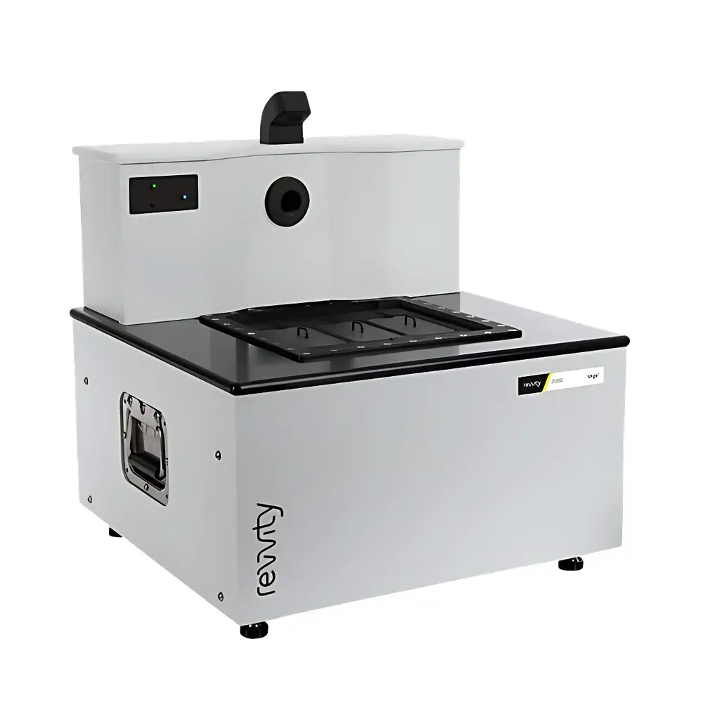
| Throughput | Up to 3 mice per imaging session |
| Field of View | Wide-Field Volumetric Scanning |
| Regulatory Status | CE-marked, FDA-cleared for preclinical research use |
Overview
The Revvity INV Vega is a next-generation, fully automated small animal ultrasound imaging system engineered for high-fidelity, reproducible preclinical in vivo studies. Unlike conventional handheld ultrasound platforms, the INV Vega implements a bottom-up, motorized transducer architecture integrated beneath a precision-engineered imaging stage. This design eliminates operator-dependent variability by removing manual probe manipulation—replacing it with programmable, repeatable transducer positioning and synchronized motion control. The system operates on clinical-grade pulse-echo ultrasound principles, utilizing high-frequency broadband transducers (center frequencies optimized for murine anatomy) to deliver real-time B-mode, M-mode, and Doppler-based imaging. Its wide-field volumetric acquisition capability enables whole-body or organ-level scanning without repositioning the subject, supporting longitudinal assessment of anatomical structure, hemodynamics, tissue stiffness, and microvascular perfusion in live mice. Designed specifically for translational research environments, the INV Vega meets the functional and regulatory expectations of GLP-compliant preclinical labs—including audit-ready data capture, metadata-rich DICOM export, and full traceability of acquisition parameters.
Key Features
- Fully automated, hands-free transducer positioning via integrated XYZ motorized stage with sub-millimeter repeatability
- Simultaneous multi-animal imaging support: accommodates up to three anesthetized mice on a single imaging platform with independent region-of-interest (ROI) targeting
- Wide-field 3D volumetric scanning: enables rapid whole-organ or systemic coverage with isotropic resolution and minimal stitching artifacts
- Dual-integrated transducer configuration: includes high-resolution linear array for superficial structures and phased array for deep-tissue penetration and cardiac imaging
- Clinically validated Shear Wave Elastography (SWE) mode: provides quantitative Young’s modulus mapping (kPa) of soft tissues with calibrated mechanical loading and wave propagation analysis
- Contrast-Enhanced Ultrasound (CEUS) with microbubble-specific pulse sequencing: supports dynamic vascular perfusion quantification using time-intensity curve analysis and destruction-replenishment kinetics
- Real-time 4D (3D + time) cine acquisition with ECG/respiratory gating compatibility for motion-compensated cardiac and respiratory cycle analysis
Sample Compatibility & Compliance
The INV Vega is validated for use with adult and juvenile C57BL/6, BALB/c, nude, and NSG mouse models (18–30 g body weight). Anesthesia integration supports isoflurane delivery via nose cone with built-in physiological monitoring (temperature, respiration rate, SpO₂). All hardware and software comply with IEC 62304 (medical device software lifecycle), ISO 13485 (quality management), and EU MDR Annex II requirements for Class IIa preclinical imaging systems. Data integrity conforms to ALCOA+ principles; audit trails record user actions, parameter changes, and image acquisition timestamps. The system supports 21 CFR Part 11-compliant electronic signatures when deployed with Revvity’s optional LIMS-integrated software module.
Software & Data Management
Acquisition and analysis are performed through VegaView™, a dedicated Windows-based application built on Qt and CUDA-accelerated processing kernels. The software provides DICOM 3.0-compliant import/export, vendor-neutral storage via configurable PACS integration, and native support for NIfTI and MINC formats for downstream MRI/PET co-registration. Quantitative modules include ROI-based strain elastography analytics, time-intensity curve fitting (TIC) for CEUS kinetics, ejection fraction calculation from gated cardiac loops, and volumetric tumor burden segmentation using adaptive thresholding and morphological filtering. All processed datasets retain embedded acquisition metadata—including transducer ID, mechanical index (MI), thermal index (TI), frame rate, depth, and gain settings—for full experimental traceability.
Applications
- Longitudinal oncology studies: monitoring orthotopic or subcutaneous tumor growth, necrosis, and anti-angiogenic response via CEUS and SWE
- Cardiovascular phenotyping: quantifying left ventricular mass, diastolic function, and wall motion abnormalities in genetic or pharmacologically induced models
- Fibrosis progression modeling: non-invasive staging of liver, kidney, or myocardial stiffness changes across disease timelines
- Immunotherapy response assessment: correlating microvascular remodeling and stromal stiffness with T-cell infiltration patterns
- Developmental biology: high-throughput embryonic and postnatal structural screening in transgenic lines
- Toxicology endpoints: detecting early echogenicity shifts, chamber dilation, or perfusion deficits following compound exposure
FAQ
Is the INV Vega compatible with existing Revvity imaging workflows?
Yes—the system shares common data architecture and metadata schema with Revvity’s IVIS SpectrumCT and Quantum FX platforms, enabling cross-modality analysis within the same informatics environment.
Does the system require specialized sonographer training?
No—VegaView™ features guided workflow templates and context-aware tooltips; standard operation requires ≤2 hours of hands-on orientation for experienced rodent researchers.
Can raw RF data be exported for custom signal processing?
Yes—proprietary .vegaRF binary format is documented in the SDK; MATLAB and Python APIs are available under Revvity’s Research Collaboration License.
What is the maximum imaging depth achievable in murine abdominal scans?
Up to 25 mm at 15 MHz center frequency with maintained axial resolution < 120 µm and lateral resolution < 200 µm (ISO 10942-1 compliant measurement).
How does the system handle motion artifacts during free-breathing acquisitions?
Integrated respiratory gating uses real-time plethysmographic feedback; optional ECG synchronization supports cardiac-phase binning with temporal resolution down to 5 ms per frame.


