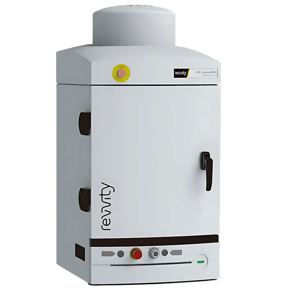
Revvity IVIS Lumina XRMS Series III Small Animal In Vivo Imaging System
| Brand | Revvity |
|---|---|
| Origin | USA |
| Manufacturer | Revvity, Inc. |
| Product Type | Imported |
| Model | IVIS Lumina XRMS Series III |
| Imaging Modalities | Bioluminescence, Multispectral Fluorescence, Radioisotope (Cerenkov), X-ray |
| Animal Capacity | Mice and Rats (up to 600 g) |
| Optical Filter Set | 26-position automated filter wheel |
| X-ray Detector | High-resolution, low-dose CMOS-based digital radiography system |
| Software Platform | Living Image® 4.7+ with DyCE™ module support |
| Regulatory Compliance | Designed for GLP-compliant workflows |
Overview
The Revvity IVIS® Lumina XRMS Series III is a high-performance, two-dimensional multimodal in vivo imaging platform engineered for preclinical research requiring simultaneous functional and anatomical visualization in small laboratory animals. Built upon the proven IVIS optical imaging architecture, this third-generation system integrates four complementary imaging modalities—bioluminescence, multispectral fluorescence, Cerenkov radiation (for radioisotope detection), and low-dose X-ray—within a single, unified instrument. Its core measurement principle relies on photon detection via a back-illuminated, thermoelectrically cooled CCD camera (1024 × 1024 pixels, >95% quantum efficiency at 600 nm), coupled with precision spectral filtering and calibrated X-ray projection geometry. Unlike conventional optical-only systems, the Lumina XRMS III enables spatial registration of functional signals (e.g., luciferase activity or fluorophore distribution) onto high-fidelity anatomical reference maps derived from X-ray radiography—eliminating reliance on external CT or MRI co-registration. This capability is particularly critical in longitudinal studies where consistent anatomical referencing across timepoints ensures quantitative rigor in tumor burden assessment, infection localization, or graft viability monitoring.
Key Features
- Integrated multimodal acquisition: Simultaneous or sequential bioluminescence, multispectral fluorescence (400–900 nm), Cerenkov, and X-ray imaging within one imaging session.
- 26-position automated filter wheel: Enables precise spectral unmixing of up to 12 fluorescent probes in a single acquisition using linear unmixing algorithms embedded in Living Image® software.
- Dual-mode X-ray subsystem: Configurable field-of-view (FOV) optimized for mice (up to 30 cm × 25 cm) and rats (up to 40 cm × 30 cm); uses a CsI:Tl scintillator coupled to a high-sensitivity CMOS detector (pixel size: 100 µm, spatial resolution: ≤50 µm at 1× magnification).
- Optical-X-ray co-registration engine: Proprietary geometric calibration ensures sub-millimeter alignment accuracy between optical and X-ray image planes across all FOVs—no manual overlay or post-hoc registration required.
- Thermally stabilized optical chamber: Maintains ambient temperature control (±0.5°C) during long exposures to minimize thermal noise and ensure signal reproducibility.
- Factory-calibrated photometric standardization: Each instrument undergoes NIST-traceable optical throughput characterization; enables cross-platform data comparability in multi-site studies.
Sample Compatibility & Compliance
The IVIS Lumina XRMS III supports live imaging of rodents—including mice (typically 15–35 g), rats (200–600 g), and other small mammals such as hamsters and guinea pigs—under controlled anesthesia (isoflurane-compatible stage with integrated gas delivery ports). The system accommodates standard animal positioning accessories (e.g., prone/supine holders, tail-vein injection trays) and is compatible with common contrast agents including barium sulfate (GI tract opacification), iodinated compounds (vascular imaging), and clinically relevant radiotracers (e.g., 18F-FDG, 99mTc-MDP). From a regulatory standpoint, the hardware and Living Image® software are designed to support Good Laboratory Practice (GLP) and Good Manufacturing Practice (GMP)-aligned workflows. Optional 21 CFR Part 11 compliance packages include electronic signature enforcement, role-based access control, and immutable audit trails for all acquisition and analysis events—facilitating FDA submission readiness.
Software & Data Management
Living Image® software (v4.7 or later) serves as the central interface for instrument control, image acquisition, spectral unmixing, quantification, and report generation. Its modular architecture includes: (1) an Imaging Wizard that guides users through modality-specific acquisition protocols; (2) Region-of-Interest (ROI) tools with background subtraction, auto-thresholding, and kinetic curve extraction; (3) Dynamic Contrast Enhancement (DyCE™) module for time-resolved pharmacokinetic modeling of probe distribution; and (4) export modules compliant with MIAME and MINC standards for integration into institutional LIMS or ELN systems. All raw image data (TIFF/FITS format) and metadata—including exposure time, binning, f-stop, filter position, X-ray kV/mAs settings—are embedded in EXIF headers. Data encryption, DICOM-SR export, and secure LDAP/Active Directory authentication are available via enterprise licensing.
Applications
This platform is extensively deployed in oncology (orthotopic/metastatic tumor tracking, therapy response evaluation), infectious disease (bacterial/fungal/viral load quantification, biofilm formation), immunology (cell trafficking, inflammation kinetics), and regenerative medicine (stem cell engraftment, tissue scaffold integration). Published use cases include longitudinal monitoring of 4T1-luc2 mammary carcinoma bone metastasis with concurrent X-ray–confirmed osteolysis, real-time gastrointestinal transit mapping using dual-labeled Salmonella Xen26-luxcherry under barium-enhanced radiography, and chronic periprosthetic joint infection surveillance via combined near-infrared fluorescence (FolateRSenseTM680) and skeletal X-ray fusion. Its ability to resolve anatomically localized functional changes makes it indispensable for hypothesis-driven mechanistic studies requiring spatial context beyond whole-body signal intensity.
FAQ
Does the IVIS Lumina XRMS III require external shielding or dedicated room installation?
No. The X-ray subsystem operates at ≤40 kV and delivers ≤0.5 mGy per exposure—well below occupational exposure limits—allowing safe operation in standard vivarium or core facility environments without structural modifications.
Can Living Image® software perform absolute quantification across instruments?
Yes. Factory-applied photonic calibration coefficients enable normalization of radiance values (p/sec/cm2/sr) to physical photon flux, supporting inter-instrument and inter-laboratory quantitative comparisons when acquisition parameters are matched.
Is DyCE™ compatible with non-optical tracers such as PET or SPECT isotopes?
DyCE™ is specifically optimized for optical reporters (bioluminescent enzymes, fluorescent dyes, Cerenkov emitters). It does not reconstruct tomographic data or replace nuclear imaging modalities but provides complementary 2D dynamic biodistribution analysis where optical sensitivity and temporal resolution are prioritized.
What maintenance is required for the X-ray subsystem?
Annual performance verification by Revvity-certified service engineers is recommended, including detector gain calibration, X-ray tube output validation, and mechanical alignment checks—all documented in accordance with ISO 13485 quality system requirements.
Are there validated SOPs available for GLP-compliant study execution?
Revvity provides a comprehensive set of instrument-specific SOP templates covering daily operational checks, preventive maintenance, software validation, and data integrity practices—fully adaptable to sponsor or regulatory authority requirements.


