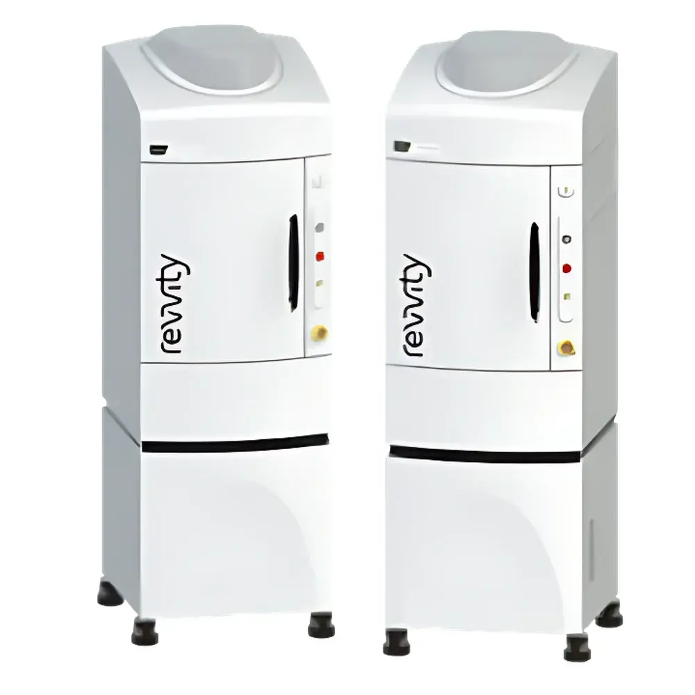
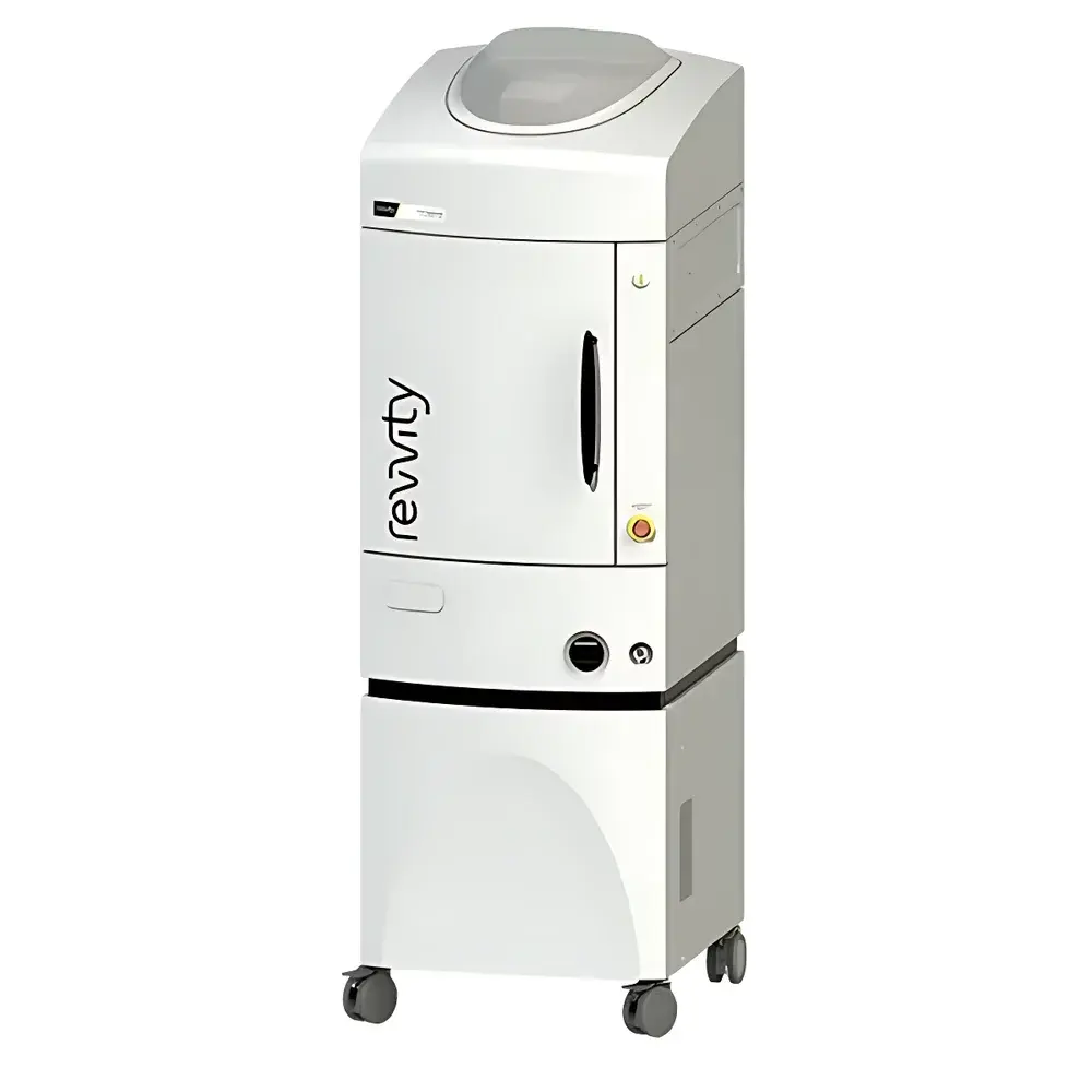
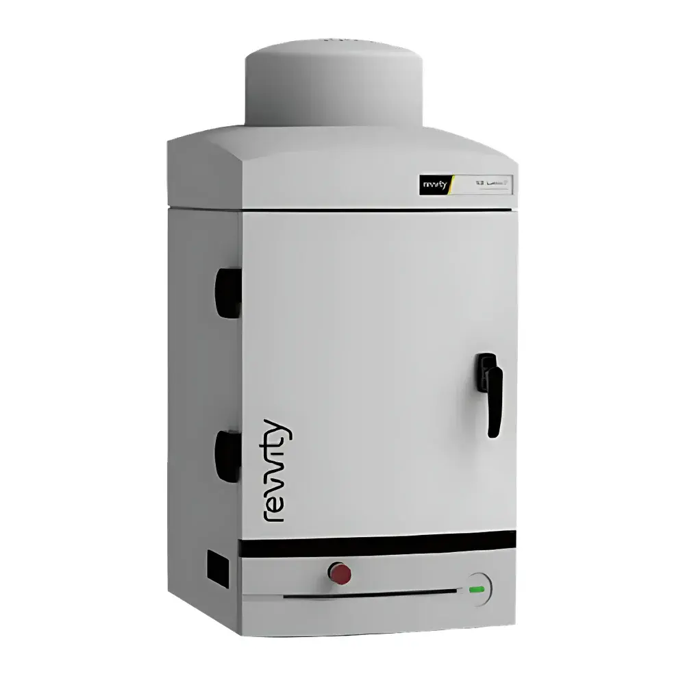
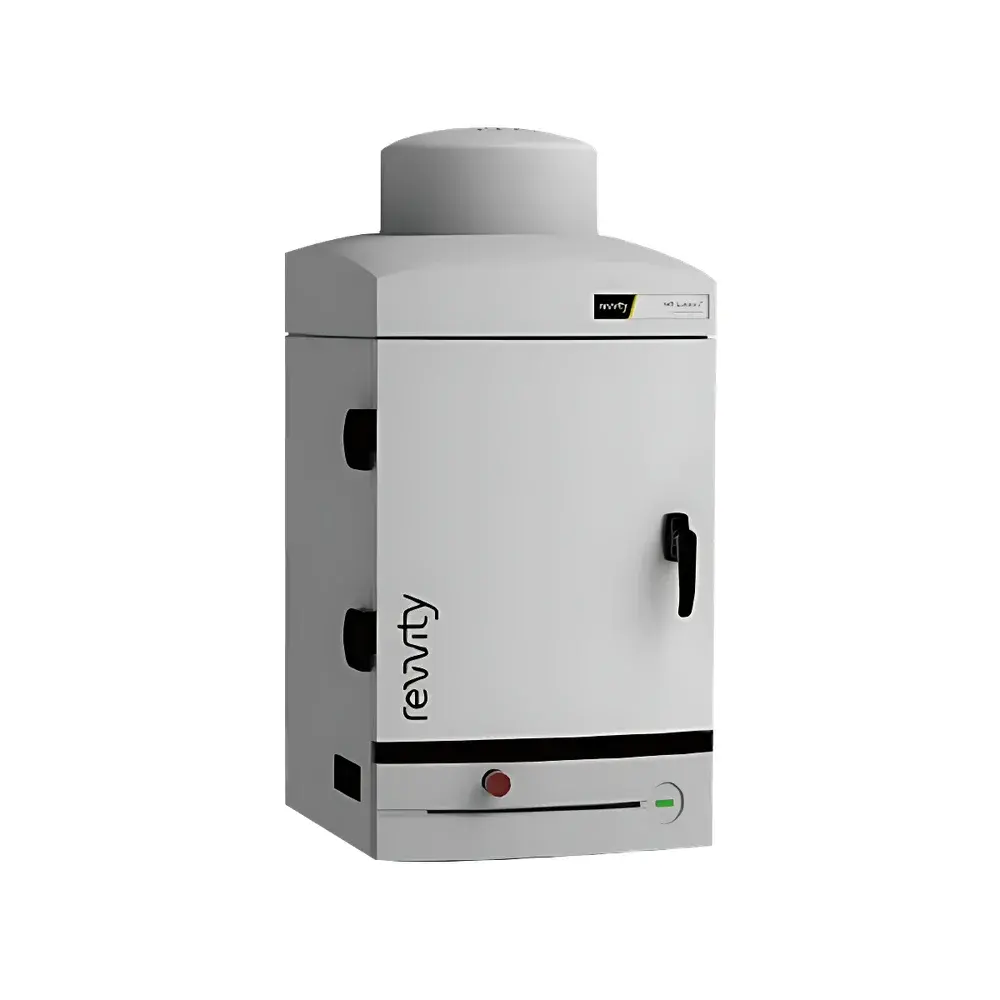
Revvity IVIS Spectrum CT2 and IVIS Lumina Series Small Animal In Vivo Imaging Systems
| Brand | Revvity |
|---|---|
| Origin | USA |
| Manufacturer Type | Original Equipment Manufacturer (OEM) |
| Product Category | Imported Instrument |
| Model(s) | IVIS Spectrum CT2, IVIS Lumina LT / Series III / XRMS Series III / S5 / X5 / Spectrum2 |
| Imaging Modality | Optical (Bioluminescence, Fluorescence, Cerenkov), X-ray, microCT, Ultrasound |
| Animal Models | Mouse, Rat, Rabbit |
| Regulatory Compliance | FDA-cleared for preclinical research use |
Overview
The Revvity IVIS® Spectrum CT2 and IVIS Lumina® series represent a modular platform of preclinical in vivo imaging systems engineered for quantitative, multimodal visualization of biological processes in live small animals. These systems integrate core optical detection principles—including photon-counting intensified CCD (ICCD) technology for bioluminescence imaging (BLI), spectral unmixing algorithms for fluorescence molecular tomography (FMT)-compatible multispectral fluorescence imaging (MSFI), and Cerenkov luminescence imaging (CLI) for radiotracer visualization—within a thermally stabilized, light-tight chamber. The IVIS Spectrum CT2 extends this capability by co-registering high-resolution optical data with low-dose microcomputed tomography (microCT) volumetric anatomy, enabling true 3D anatomical-functional fusion. All models operate under controlled isoflurane anesthesia delivery and temperature-regulated animal staging, ensuring physiological consistency across longitudinal studies.
Key Features
- High-sensitivity ICCD detection with quantum efficiency >35% at 500–700 nm and dark current <0.001 e−/pixel/sec at −90°C cooling
- Automated spectral acquisition using up to 28 precision bandpass filters (430–850 nm) for robust autofluorescence subtraction and multiplexed probe quantification
- Integrated microCT subsystem (IVIS Spectrum CT2 only): isotropic spatial resolution down to 45 µm, dose-efficient scanning (<5 mGy per scan), and reconstruction-based attenuation correction for quantitative optical signal normalization
- High-throughput configurations (IVIS Lumina S5/X5): simultaneous imaging of up to five mice with independent positioning and real-time motion compensation
- X-ray imaging module (IVIS Lumina XRMS/X5): 50 kVp source, 10 µm focal spot, and digital detector enabling high-contrast skeletal and soft-tissue visualization with effective dose ≤1.2 mGy per exposure
- Standardized calibration protocols traceable to NIST-certified light sources and radiometric standards for cross-instrument reproducibility
Sample Compatibility & Compliance
The platform supports longitudinal imaging of immunocompetent and immunodeficient murine models (C57BL/6, BALB/c, NSG), Sprague-Dawley and Wistar rats, and New Zealand White rabbits. Imaging workflows comply with NIH Guide for the Care and Use of Laboratory Animals and AAALAC International standards. Data acquisition and storage adhere to ALCOA+ principles (Attributable, Legible, Contemporaneous, Original, Accurate, Complete, Consistent, Enduring, Available). Software audit trails meet FDA 21 CFR Part 11 requirements for electronic records and signatures in regulated environments. System validation documentation includes IQ/OQ/PQ protocols aligned with ISO/IEC 17025 and ASTM E2500-13 for analytical instrument qualification.
Software & Data Management
Living Image® 4.7 software provides a unified interface for acquisition, spectral unmixing, 3D reconstruction (Spectrum2/Spectrum CT2), and co-registration with microCT or ultrasound datasets. It supports batch processing, ROI-based kinetic analysis, and export to DICOM, NIfTI, and MATLAB-compatible formats. Built-in pharmacokinetic modeling tools enable compartmental analysis of tracer distribution. All processing steps are scriptable via Python API, facilitating integration into automated pipelines compliant with FAIR (Findable, Accessible, Interoperable, Reusable) data principles. Raw image metadata—including exposure time, binning, filter set, anesthesia status, and animal ID—is embedded in TIFF headers and archived with SHA-256 checksums.
Applications
- Oncology: Tumor growth kinetics, metastatic burden quantification, and therapeutic response monitoring using luciferase-expressing xenografts or NIR fluorescent probes
- Immunology: Tracking adoptively transferred immune cells labeled with near-infrared dyes or luciferase reporters
- Cardiovascular research: Assessment of myocardial infarction remodeling via CLI with 64Cu-PTSM or 89Zr-labeled antibodies
- Neuroscience: Visualizing neuroinflammation with TSPO-targeted fluorescent tracers and correlating signal intensity with microCT-derived ventricular volume changes
- Drug development: Validating target engagement and biodistribution of novel radiolabeled therapeutics under GLP-compliant study conditions
- Regenerative medicine: Monitoring stem cell engraftment and differentiation using dual-reporter constructs (e.g., firefly luciferase + tdTomato)
FAQ
What regulatory standards does the IVIS platform support for GxP-compliant studies?
The system supports 21 CFR Part 11 compliance through electronic signature enforcement, immutable audit trails, and role-based access control in Living Image software. Installation and operational qualification packages are available for GLP and GCP environments.
Can IVIS Spectrum CT2 perform simultaneous optical and microCT acquisition?
No—optical and microCT modalities are acquired sequentially but automatically co-registered using fiducial markers and iterative closest point (ICP) algorithms. Total session time for combined acquisition is typically under 15 minutes per animal.
Is spectral unmixing validated for more than three fluorescent probes?
Yes. Validation data demonstrate linear separation of up to five spectrally distinct probes (e.g., Cy5.5, IRDye800CW, Alexa Fluor 750, DY-731, and CF790) using the 28-filter acquisition protocol and constrained non-negative matrix factorization (cNMF) algorithms.
How is radiation dose managed during microCT imaging?
Dose modulation is implemented via automatic exposure control (AEC), pulsed X-ray emission, and hardware collimation. Dose maps are generated per scan and logged alongside raw projections to ensure ALARA (As Low As Reasonably Achievable) compliance.
Does the platform support third-party probe characterization?
Yes. The system accepts user-defined excitation/emission spectra and quantum yield inputs for custom probe libraries, enabling accurate in vivo concentration estimation using calibrated phantom-based reference standards.
Related Products


