FluorCam Open-Field Chlorophyll Fluorescence Imaging System by PSI
| Brand | PSI (PhotoSystems Instruments) |
|---|---|
| Origin | Czech Republic |
| Model | FluorCam Open-Field Chlorophyll Fluorescence Imaging System |
| Imaging Area | Standard 13 × 13 cm |
| CCD Sensor | TOMI-2, 1360 × 1024 px, 16-bit A/D, 6.45 µm pixel size, 20 fps max |
| Light Sources | 4 × LED panels (617 nm measuring light |
| dual actinic | 617 nm + white or 617 nm + 470 nm |
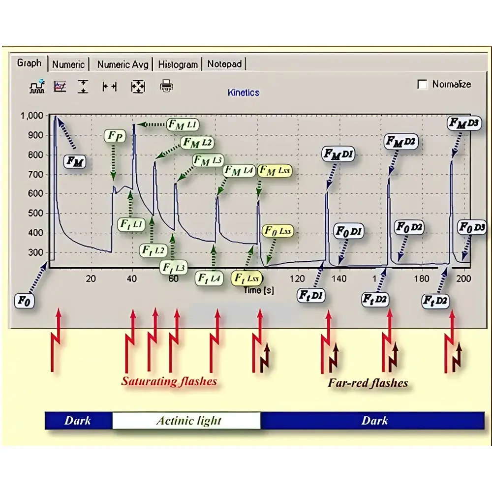
| Fluorescence Parameters | >50 including Fo, Fm, Fv/Fm, ΦPSII, NPQ, qL, ETR, Rfd, QY_Ln, FtDn, etc. |
| Software | FluorCam v8.x with protocol scripting, ROI analysis (>1000 regions), timestamped automated acquisition, dual averaging modes, export to Excel & video |
| Optional Modules | TetraCam RGB imaging (20 × 25 cm), PAR/NDVI (735 nm + 650 nm), GFP/YFP/DAPI imaging (with 7-position filter wheel & UV/blue/green LED panels), multi-spectral fluorescence capability |
Overview
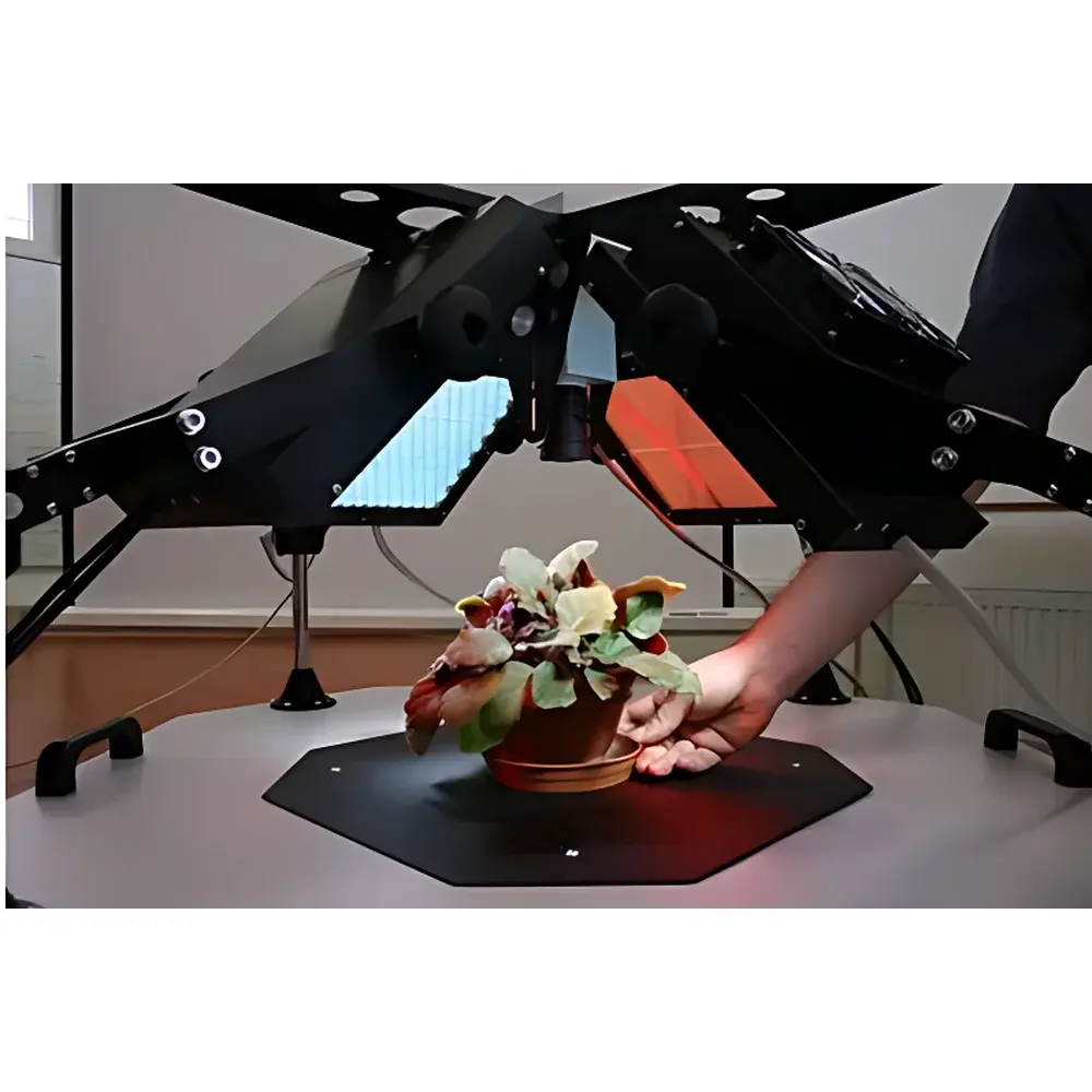
The FluorCam Open-Field Chlorophyll Fluorescence Imaging System by PSI is a high-resolution, modular platform engineered for non-invasive, quantitative assessment of photosynthetic performance across spatial and temporal scales. Based on the biophysical principles of chlorophyll a fluorescence induction kinetics—governed by the photochemical and non-photochemical quenching pathways of Photosystem II—the system captures dynamic fluorescence responses under precisely controlled light regimes. Unlike conventional single-point fluorometers, FluorCam delivers two-dimensional parameter maps (e.g., ΦPSII, NPQ, Fv/Fm) at pixel-level resolution, enabling spatial heterogeneity analysis across leaves, whole plants, algae cultures, mosses, lichens, or multi-well plates. Its open-field architecture eliminates optical enclosure constraints, permitting direct integration with growth chambers, climate rooms, or greenhouse environments—critical for longitudinal stress phenotyping and ecophysiological field simulation.
Key Features
- Modular optical design: Four independently adjustable LED panels (standard 617 nm red measuring light; dual actinic sources configurable as red+white or red+blue; saturating flash up to 4000 µmol·m⁻²·s⁻¹) support standardized protocols (Kautsky induction, OJIP, light-response curves, fluorescence quenching analysis) and custom irradiance sequences.
- High-fidelity imaging: TOMI-2 scientific CCD sensor with 1360 × 1024 resolution, 16-bit dynamic range (65,536 gray levels), 6.45 µm pixel pitch, and real-time acquisition at up to 20 frames per second—optimized for both kinetic fluorescence transients and static snapshot imaging of GFP, YFP, DAPI, or wtGFP.
- Scalable field-of-view: Standard configuration supports 13 × 13 cm imaging area; large-format variant extends to 20 × 20 cm—sufficient for intact Arabidopsis rosettes, cereal seedlings, or multi-plant arrays in 96-/384-well plates.
- Extensible spectral capability: Optional LED modules (365/385 nm UV, 470 nm blue, 530 nm green, 627–740 nm far-red) combined with a motorized 7-position filter wheel enable multi-parametric fluorescence imaging—including PAR absorption, NDVI, and multi-color protein reporter quantification.
- Automated experimental control: Protocol-driven acquisition allows fully unattended operation—time-stamped datasets are stored with metadata (light intensity, duration, spectral composition) compliant with FAIR data principles. Custom protocols are scripted via an embedded language supporting nested loops, conditional logic, and hardware synchronization.
Sample Compatibility & Compliance
The system accommodates diverse biological specimens without sample preparation: detached or attached leaves, floral organs, fruits, stems, bryophytes, algal suspensions, soil microcosms, and high-throughput screening plates. Its open geometry permits integration with environmental control systems (e.g., CO2-regulated growth chambers, temperature-gradient tables), facilitating studies under physiologically relevant conditions. Data acquisition and storage adhere to GLP-aligned traceability requirements: all measurements include embedded timestamps, instrument configuration logs, and user-defined annotations. While not FDA 21 CFR Part 11-certified out-of-the-box, the software architecture supports audit trail implementation and electronic signature integration for regulated environments upon customer-specific validation.
Software & Data Management
FluorCam v8.x provides a unified interface for live monitoring, protocol definition, pre-processing, and quantitative analysis. Region-of-interest (ROI) selection supports arbitrary polygons, circles, sectors, or auto-segmented objects (>1000 ROIs per image). Two signal-processing modes—“signal calculation then averaging” (optimal for high-SNR kinetics) and “signal averaging then calculation” (robust against transient noise)—ensure statistical rigor across experimental conditions. Output formats include time-resolved parameter movies, false-color parameter maps, Excel-exported kinetic curves, histograms, and annotated ROI summary tables. The software exports MIAME-compliant metadata and supports batch processing for longitudinal experiments spanning days or weeks.
Applications
- Photosynthetic phenotyping: High-throughput screening of Fv/Fm, ΦPSII, and ETR for crop breeding programs and mutant libraries.
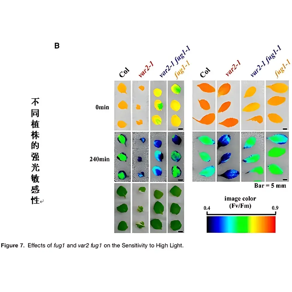
- Abiotic stress physiology: Quantifying spatial heterogeneity in drought-, heat-, salinity-, or heavy metal-induced photoinhibition and recovery dynamics.
- Stomatal function mapping: Using blue-light-induced fluorescence quenching (via optional 470 nm panel) to resolve stomatal conductance gradients across leaf surfaces.
- Plant-microbe interactions: Monitoring localized PSII inhibition during pathogen colonization or symbiont establishment.
- Multispectral reporter imaging: Co-localizing chlorophyll fluorescence with GFP/YFP/DAPI signals to correlate photosynthetic status with gene expression or cellular integrity.
- Ecotoxicology: Assessing sub-lethal effects of pollutants (e.g., Cd, Pb, herbicides) on photosynthetic efficiency in bioindicators such as Lemna or Chlamydomonas.
FAQ
What is the minimum detectable change in Fv/Fm that FluorCam can reliably resolve?
Under standard measurement conditions (25°C, dark-adapted samples), the system achieves a typical Fv/Fm precision of ±0.008 (CV < 1.2%) across replicate pixels—sufficient to detect subtle physiological shifts induced by early-stage stress.
Can FluorCam be synchronized with external environmental controllers?
Yes—via TTL triggers and Ethernet-based API commands, the system integrates with programmable climate chambers, irrigation systems, and gas analyzers for closed-loop stress imposition and response capture.
Is calibration required before each experiment?
No routine recalibration is needed; the LED output stability is factory-characterized and drift-compensated in software. Users may perform optional daily uniformity checks using reference standards.
Does the software support batch analysis of time-series datasets?
Yes—batch processing scripts automate ROI extraction, parameter computation, and statistical comparison across hundreds of time points, with output structured for downstream analysis in R, Python, or MATLAB.
What safety certifications does the system meet?
The FluorCam platform complies with CE, RoHS, and IEC 61000-6-3 (EMC) standards. LED panels operate within Class 1 laser safety limits per IEC 60825-1:2014.


