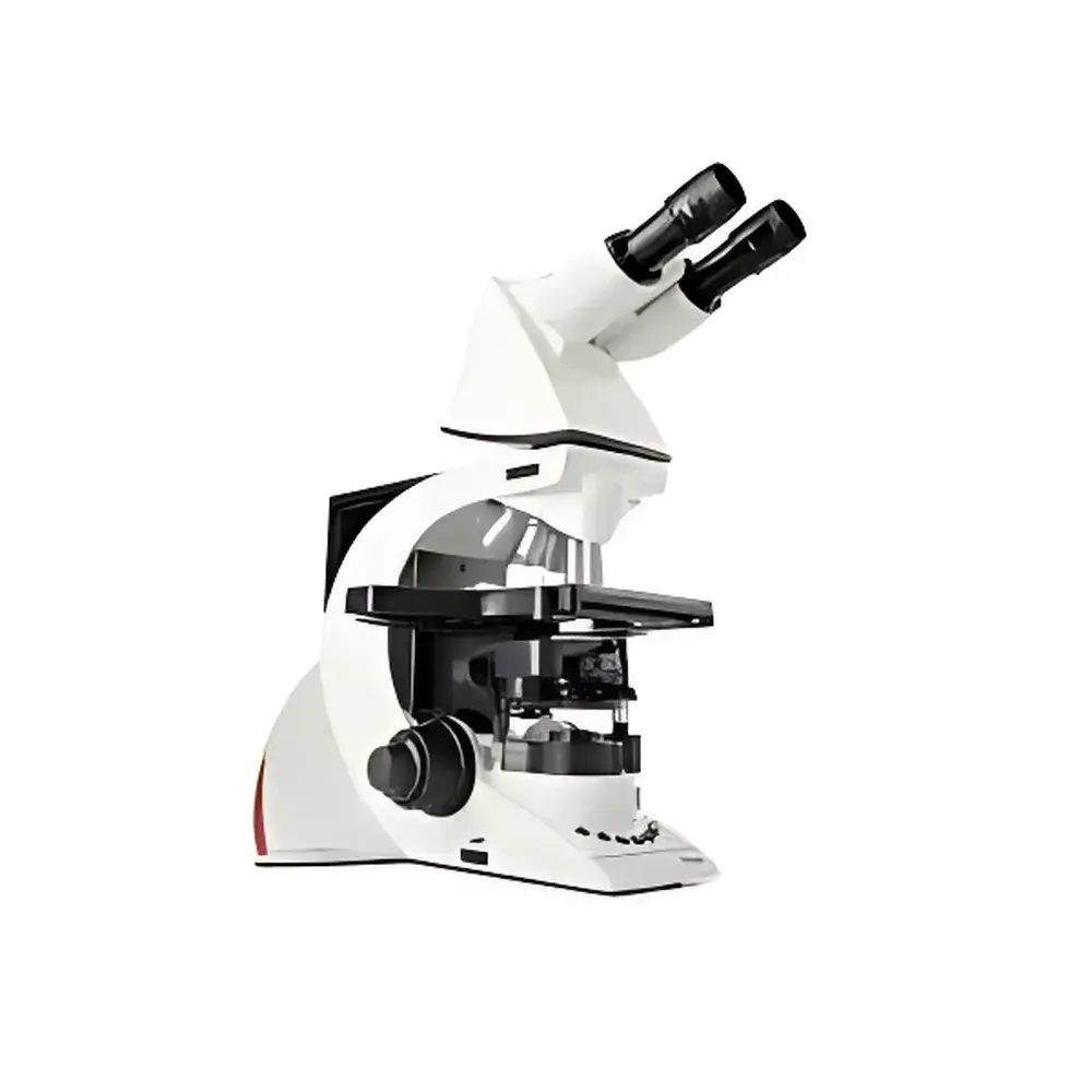
Leica DM3000 Biological Microscope
| Brand | Leica |
|---|---|
| Country of Origin | Germany |
| Microscope Type | Upright |
| Eyepiece Configuration | Binocular |
| Objective Turret | Motorized 6-Position |
| Condenser | Automated High-Aperture (0.90 NA) with Position Memory |
| Illumination | Motorized Brightness Control per Objective |
| Ergonomic Features | Adjustable-Height Focus Knobs (Patent DE10340721), Symmetrical XY Stage, One-Hand Operation, ErgoStage with Ceramic Surface |
| Software Compatibility | Leica Application Suite (LAS) for Pathology & Cytology Imaging |
| Compliance | Designed for ISO 13485-aligned clinical laboratories, supports GLP/GMP documentation workflows via LAS audit trail and user access control |
Overview
The Leica DM3000 is a high-performance upright biological microscope engineered for routine diagnostic and research applications in pathology, cytology, hematology, and biomedical laboratories. Built on Leica Microsystems’ proven optical platform, it employs Köhler illumination principles with motorized condenser positioning and intensity regulation to ensure consistent, reproducible image quality across magnifications. Its core architecture integrates precision mechanical design with intelligent automation—enabling rapid transitions between user-defined objectives, adaptive light management, and ergonomic operability without compromising optical fidelity. Unlike manually adjusted conventional microscopes, the DM3000’s closed-loop optical alignment system maintains optimal illumination geometry for each objective, minimizing user-induced variability and supporting standardized workflow protocols required in regulated clinical environments.
Key Features
- Motorized 6-position objective turret: Switches between objectives in under 0.5 seconds; supports programmable dual-objective “toggle mode” for rapid comparison (e.g., 10× vs. 40× during cytological screening).
- Automated condenser head (0.90 NA): Moves to pre-calibrated height positions synchronized with objective magnification and numerical aperture—ensuring optimal resolution and contrast without manual recentering or refocusing.
- Dynamic brightness control: Adjusts halogen lamp intensity based on selected objective transmission characteristics, maintaining constant perceived brightness and reducing photobleaching in stained specimens.
- Ergonomic focus system: Patented adjustable-height focus knobs (DE10340721) allow vertical positioning to match operator anthropometry; symmetrical XY stage controls and one-hand operation reduce repetitive strain during prolonged examination sessions.
- Ceramic-coated mechanical stage (ErgoStage): Provides wear-resistant, low-friction movement with precise X/Y repeatability; compatible with standard slide holders and one-handed slide exchange mechanisms.
- Modular optical train: Accepts trinocular phototube (HC L1T 4/5/7), fluorescence modules, phase contrast rings (UCA S1), and daylight filters—all aligned to ISO 8036/1 and DIN standards for traceable calibration.
Sample Compatibility & Compliance
The DM3000 accommodates standard 1×3 inch (25×75 mm) glass slides and 22×22 mm coverslips, supporting brightfield, phase contrast, and optional fluorescence imaging with Leica’s high-transmission filter sets. Its optical path is optimized for routine H&E, Papanicolaou, Giemsa, and immunohistochemical staining protocols. The system complies with IEC 61000-6-3 (EMC) and IEC 61000-6-2 (immunity) standards. When paired with Leica Application Suite (LAS) software, it supports audit-trail-enabled documentation compliant with FDA 21 CFR Part 11 requirements for electronic records and signatures—making it suitable for GLP- and GMP-regulated pathology labs conducting QC/QA assessments.
Software & Data Management
The microscope interfaces seamlessly with Leica Application Suite (LAS) v4.x or later, providing integrated acquisition, annotation, measurement, and reporting tools tailored for cytology and histopathology. LAS enables multi-user role-based access control, timestamped image metadata logging, and export to DICOM-compliant archives. Optional LAS X Core modules support automated cell counting, mitotic index quantification, and region-of-interest (ROI) batch analysis. All system configurations—including turret position, condenser height, and illumination settings—are stored per user profile and recalled automatically upon login, ensuring procedural consistency across shifts.
Applications
Primary use cases include cervical cytology screening (Bethesda System), bone marrow morphology assessment, peripheral blood smear evaluation, frozen section analysis, and routine histological diagnosis. Its stable mechanical platform and thermal-compensated focus drive minimize drift during extended observation—critical for time-sensitive intraoperative consultations. In academic research, the DM3000 serves as a reliable platform for teaching microscopy fundamentals, comparative morphological studies, and method validation prior to deployment of advanced imaging systems.
FAQ
Is the DM3000 compatible with digital camera systems?
Yes—it supports Leica DFC series cameras via C-mount interface and offers full hardware synchronization with LAS for live preview, capture, and metadata embedding.
Can the motorized functions be disabled for manual operation?
All motorized components (turret, condenser, illumination) can be operated manually using dedicated override levers without requiring service intervention.
Does the system support phase contrast out-of-the-box?
Phase contrast capability requires optional UCA S1 condenser bottom unit and matching phase rings; these are available as factory-configured bundles or field-upgradable kits.
What lamp type is used, and how is replacement performed?
It uses a standardized 12 V / 30 W halogen lamp housed in a drawer-mounted socket—accessible without tools and replaceable in under 60 seconds.
Is service and calibration support available globally?
Leica Microsystems provides authorized service network coverage in over 120 countries, including on-site preventive maintenance, optical alignment certification, and ISO/IEC 17025 traceable calibration reports.


