Leica DMi8 Inverted Research Microscope
| Brand | Leica |
|---|---|
| Origin | Germany |
| Model | DMi8 |
| Instrument Type | Inverted Fluorescence Microscope |
| Excitation Source | Metal Halide Lamp |
| Eyepiece | 10× |
| Objectives | Plan Achromat: 5×/0.1, 10×/0.25, 50×/0.7 |
| Fluorescence Module | Integrated 5-position Filter Turret with Fine Adjustment |
| Light Sources | 100 W Mercury Arc Lamp, 1200 W Long-Life Metal Halide Lamp |
| Observation Head | Trinocular |
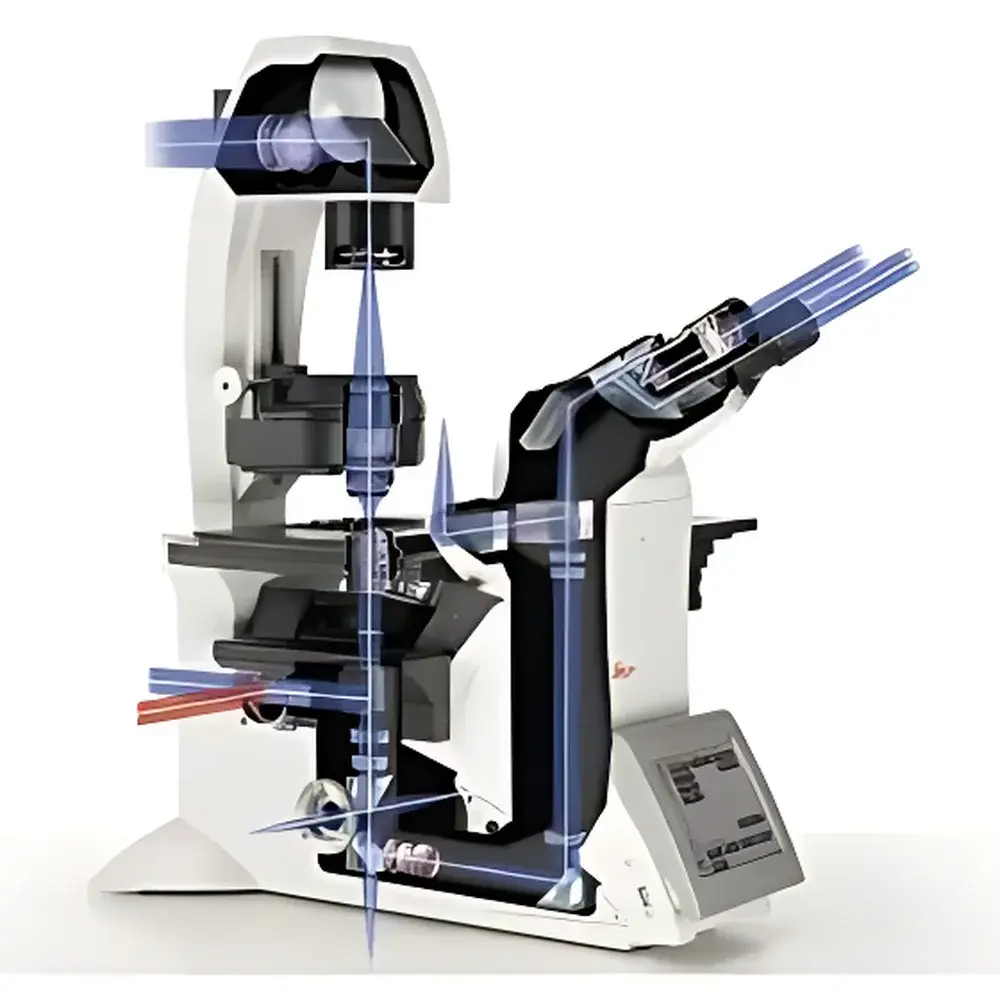
| Optical Path | Dual Infinity-Corrected Port Architecture |
Overview
The Leica DMi8 is an inverted research-grade microscope engineered for rigorous life science applications—particularly live-cell imaging, quantitative fluorescence analysis, and multimodal correlative microscopy. Built upon Leica Microsystems’ proven optical architecture, the DMi8 employs a dual infinity-corrected optical path design, enabling simultaneous access to two independent parallel light paths without optical coupling loss. This architecture supports advanced techniques including FRAP (Fluorescence Recovery After Photobleaching), optogenetic stimulation, laser ablation, and single-molecule photomanipulation—all while maintaining Köhler illumination integrity and chromatic fidelity across visible and near-UV excitation bands. Its rigid, thermally stable mechanical platform minimizes stage drift during time-lapse acquisitions, and its modular base accommodates environmental control chambers, motorized Z-drives, and high-speed filter wheels compatible with sCMOS and EMCCD detectors.
Key Features
- Dual infinity-corrected optical ports: Enables concurrent use of widefield fluorescence and auxiliary light sources (e.g., lasers, LEDs) for photostimulation or structured illumination—no beam combiner required.
- Modular, field-upgradable architecture: Supports seamless integration of motorized components—including autofocus, encoded filter turrets, XY stages, and environmental enclosures—without disassembly or recalibration.
- Integrated 5-position fluorescence filter turret with micrometer-scale adjustment: Ensures precise alignment of excitation/emission bandpass filters for optimal signal-to-noise ratio in multicolor experiments.
- High-power illumination system: Combines a 100 W mercury arc lamp for UV-sensitive fluorophores (e.g., DAPI, Hoechst) and a 1200 W long-life metal halide lamp delivering stable, flicker-free output across 360–700 nm—ideal for prolonged live-cell imaging.
- Ergonomic trinocular observation head with 10× widefield eyepieces: Optimized for extended operator use; includes adjustable interpupillary distance, diopter compensation, and optional C-mount or F-mount camera adapters.
- Plan achromat objective suite (5×/0.1, 10×/0.25, 50×/0.7): Designed for flat-field correction and minimal spherical aberration in aqueous and oil-immersion media—validated for compatibility with standard tissue culture vessels (e.g., 35 mm dishes, chambered coverslips).
Sample Compatibility & Compliance
The Leica DMi8 is routinely deployed in GLP-compliant cell biology labs and academic core facilities performing ISO/IEC 17025-aligned assays. Its optical train meets ISO 8578:2017 (microscope performance testing standards) and supports traceable calibration via NIST-traceable stage micrometers and fluorescence intensity standards. The system integrates seamlessly with commercial incubation chambers (e.g., Tokai Hit INU series) maintaining CO₂, humidity, and temperature control during acquisition—enabling longitudinal studies compliant with FDA 21 CFR Part 11 requirements when paired with LAS X software’s audit trail and electronic signature modules. All optical components are certified free of hazardous substances per RoHS Directive 2011/65/EU.
Software & Data Management
Controlled exclusively via Leica LAS X software (v3.7+), the DMi8 supports fully automated acquisition workflows—including multi-dimensional time-lapse (XYZT), spectral unmixing, deconvolution, and real-time focus maintenance using hardware-based focus lock. LAS X implements role-based user permissions, encrypted project storage, and full audit logging compliant with GxP data integrity principles. Raw image data is stored in open TIFF or OME-TIFF format, ensuring interoperability with ImageJ/Fiji, Python-based analysis pipelines (e.g., napari, scikit-image), and institutional LIMS systems. Exported metadata includes complete instrument configuration logs (objective ID, filter position, exposure time, lamp intensity), facilitating reproducibility in peer-reviewed publications.
Applications
- Live-cell dynamics: Mitotic tracking, organelle motility, calcium flux imaging using ratiometric dyes (e.g., Fura-2), and membrane trafficking assays.
- Photomanipulation workflows: Targeted photoactivation of caged compounds (e.g., caged Ca²⁺, caged ATP), subcellular FRAP, and optogenetic channelrhodopsin stimulation in neuronal cultures.
- Co-culture and 3D model imaging: High-resolution monitoring of spheroids, organoids, and endothelial barrier models under physiological gas conditions.
- Quantitative immunofluorescence: Multiplexed detection of ≥4 markers using sequential staining and spectral unmixing—validated against ISO 13766-2:2020 for fluorescence assay precision.
- Correlative light-electron microscopy (CLEM): Precise coordinate mapping enabled by integrated fiducial marker registration and export of stage coordinates for TEM grid relocation.
FAQ
Is the DMi8 compatible with third-party cameras and controllers?
Yes—the DMi8 features standard USB 3.0, GigE Vision, and Camera Link interfaces. All motorized components expose vendor-neutral APIs (GenICam-compliant) for integration with custom LabVIEW or Python control environments.
Can the dual infinity port support simultaneous confocal and widefield imaging?
No—the DMi8 is a widefield platform only. However, its second infinity port enables coupling to external scanning units (e.g., Yokogawa CSU-X1, Andor Dragonfly) for spinning-disk confocal extension, provided the scanner’s input pupil matches the port’s f-number and conjugate plane specifications.
What environmental control options are validated for use with the DMi8?
Leica-certified solutions include the Tokai Hit INU series (CO₂ + temperature + humidity), OkoLab H301 (temperature-only), and PeCon TempController 2000—each with documented thermal stability profiles (< ±0.2°C over 24 h) and vibration isolation ratings.
Does LAS X software support batch processing of large timelapse datasets?
Yes—LAS X Batch Processor enables GPU-accelerated background subtraction, flat-field correction, and maximum-intensity projection across thousands of Z-stacks, with progress monitoring and error logging for unattended overnight execution.
How is optical alignment verified after field installation?
Leica provides on-site commissioning including collimation verification using a HeNe laser reference, Köhler illumination validation with test slides, and fluorescence uniformity mapping across the FOV per ISO 9345:2019 Annex B protocols.
Related Products


