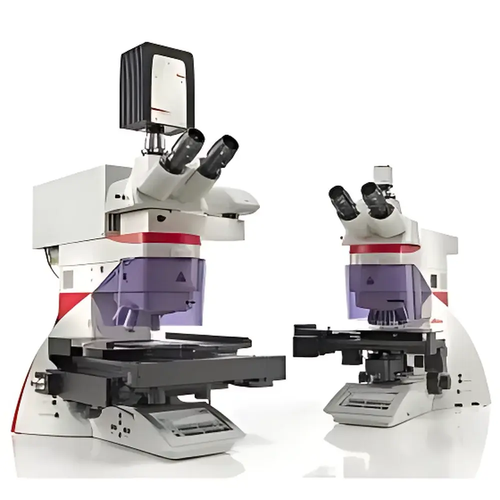
Leica LMD7 Laser Microdissection System
| Brand | Leica |
|---|---|
| Origin | Germany |
| Model | Leica LMD7 |
| Laser Wavelength | 349 nm |
| Pulse Repetition Rate | 10–5000 Hz |
| Pulse Duration | < 4 ns |
| Maximum Pulse Energy | 120 µJ |
| Optical Path | Beam-Steering via Precision Prisms (No Sample Movement) |
| Collection Method | Gravity-Based, Contact-Free, Contamination-Free |
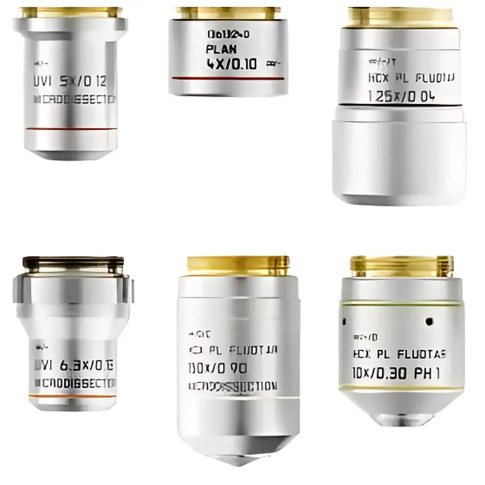
| Objective Compatibility | 10 Dry Objectives (5×–150×), Including SmartCut™ Series with UV Transmission Down to 350 nm |
| Illumination | LED (25,000 h lifetime, constant CCT) or Halogen (with CCIC) |
| Software Platform | Leica Application Suite (LAS) X v7.6 with AVC, Time-Lapse Imaging, and Workflow-Optimized Dissection Tools |
Overview
The Leica LMD7 Laser Microdissection System is a high-precision, upright optical platform engineered for contact-free, contamination-free isolation of morphologically defined regions from heterogeneous tissue sections, cytological preparations, or live-cell cultures. Unlike conventional microdissection methods that rely on mechanical transfer or sample-stage movement, the LMD7 employs a beam-steering architecture: a UV laser (349 nm) is dynamically directed across the specimen plane using motorized, high-stability prisms—leaving the sample completely stationary. This optical scanning principle ensures orthogonal, diffraction-limited laser focus perpendicular to the tissue surface, enabling clean ablation with minimal thermal spread and zero mechanical stress. The system operates under standard ambient or controlled environmental conditions (including optional climate chamber integration), and is validated for use in GLP-compliant histopathology, single-cell genomics, spatial proteomics, and developmental biology workflows where molecular integrity is non-negotiable.
Key Features
- Beam-Steering Architecture: Eliminates stage movement; laser path is precisely manipulated via calibrated prism assemblies for sub-micron positional accuracy and consistent cut geometry across large fields of view.
- UV Solid-State Laser (349 nm): Delivers up to 120 µJ pulse energy with tunable repetition rate (10–5000 Hz), enabling optimized ablation kinetics for soft tissues (e.g., brain, liver), calcified structures (bone, teeth), plant material, chromosomes, and adherent live cells.
- Gravity-Based Collection: Excised material falls freely into standard collection vessels—0.2 mL PCR tubes, 8-strip tubes, ibidi µ-Dishes, or custom microplates—ensuring no enzymatic, adhesive, or solvent-based contamination.
- SmartCut™ Objective Line: Ten dry objectives (5× to 150×) with enhanced UV transmission (down to 350 nm), optimized numerical aperture and working distance for simultaneous high-resolution imaging and precise laser targeting; 150× SmartCut objective supports sub-1 µm feature resolution in thick-sectioned samples.
- Dual Illumination Options: LED illumination (CCT-stabilized, 25,000 h lifetime, >90% energy reduction vs. halogen) or halogen with Constant Color Temperature Intelligent Control (CCIC) for stable color fidelity in brightfield, phase contrast, and fluorescence pre-screening.
- Workflow-Integrated Software (LAS X v7.6): Supports real-time laser path preview, shadow-cut mode for rapid contour ablation, time-lapse recording, automated cell recognition (AVC), and direct export of ROI coordinates to downstream NGS or mass spectrometry pipelines.
Sample Compatibility & Compliance
The LMD7 accommodates a broad spectrum of sample formats without modification: cryosections (4–20 µm), FFPE sections (1–10 µm), smears, cytospins, live-cell monolayers on PEN-membrane slides or ibidi µ-Dishes, and even intact plant leaves or insect cuticles. It complies with ISO 13485 design controls and supports audit-ready documentation per FDA 21 CFR Part 11 when configured with electronic signature and audit trail modules. All laser parameters—including pulse energy, repetition rate, spot size (via adjustable aperture), and scan velocity—are logged with timestamp and user ID. The system meets IEC 60825-1:2014 Class 4 laser safety requirements and integrates seamlessly into ISO/IEC 17025-accredited laboratories performing forensic histology, tumor microdissection for oncology biomarker validation, or single-cell RNA-seq library preparation.
Software & Data Management
Leica Application Suite (LAS) X v7.6 provides a modular, workflow-centric interface designed for routine operation by technicians and advanced configuration by bioinformaticians. Core functions include multi-layer image registration (H&E + IHC + DAPI overlays), polygonal or freehand ROI selection with pixel-level boundary refinement, batch processing of serial sections, and direct DICOM/OME-TIFF export. Optional modules include Automated Vessel Counting (AVC) for angiogenesis studies, pattern-recognition algorithms trained on tissue morphology databases, and API-driven integration with LIMS platforms (e.g., LabVantage, STARLIMS). All dissection events are stored with full metadata: laser settings, objective used, stage coordinates, operator ID, and acquisition timestamps—enabling full traceability from slide to sequencing run.
Applications
- Cancer Research: Isolation of pure tumor epithelium, stromal compartments, or circulating tumor microemboli for mutational profiling and clonal evolution analysis.
- Neuroscience: Subregion-specific dissection of hippocampal layers, cortical laminae, or dopaminergic nuclei from post-mortem human or rodent brain sections.
- Plant Biology: Targeted excision of guard cells, vascular bundles, or root cap meristems for metabolomic fingerprinting under native hydration states.
- Developmental Biology: Single-cell or small-group isolation from live zebrafish or Drosophila embryos cultured on membrane slides for scRNA-seq and lineage tracing.
- Forensic Histopathology: Recovery of trace epithelial cells from mixed biological evidence while preserving nuclear DNA integrity for STR amplification.
- Spatial Omics: Integration with MALDI-TOF or DESI-MS platforms via direct transfer onto conductive target plates for label-free metabolite mapping.
FAQ
How does gravity-based collection ensure molecular integrity?
Gravity collection eliminates contact with adhesives, solvents, or electrostatic surfaces—preserving native protein conformation, RNA integrity (RIN > 9.0 routinely achieved), and epigenetic marks such as histone modifications.
Can the LMD7 be used for live-cell isolation without compromising viability?
Yes—when combined with climate control (37°C, 5% CO₂), low-pulse-energy settings (95% post-isolation viability is maintained in adherent mammalian lines (e.g., HeLa, MCF-7) and primary neurons.
What slide types are compatible with proteomics-grade workflows?
PET membrane slides (low-additive formulation) and DIRECTOR glass slides (film-free ablation) are certified for bottom-up proteomics and untargeted metabolomics, with detection limits comparable to bulk tissue lysates.
Is remote operation supported for core facility deployment?
Yes—LAS X supports secure RDP and web-based client access with role-based permissions, enabling centralized instrument management across multi-user academic or CRO environments.
Does the system support regulatory submissions for IVD assay development?
Full validation documentation (IQ/OQ/PQ protocols), 21 CFR Part 11 compliance packages, and GMP-aligned SOP templates are available upon request for diagnostic assay qualification under CLIA or CE-IVDR frameworks.


