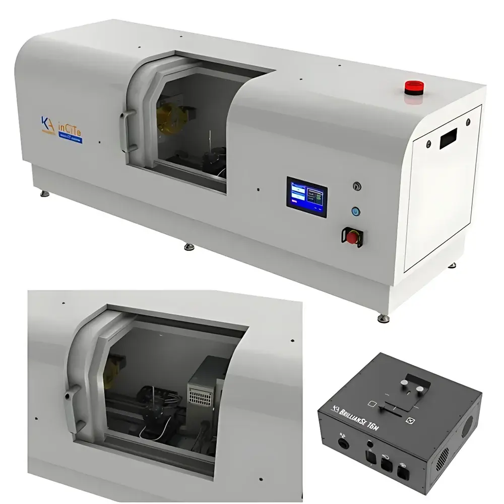
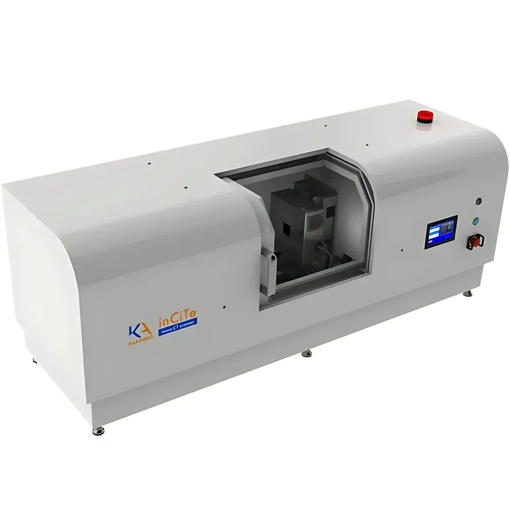
KA Imaging inCiTe Desktop Phase-Contrast MicroCT System (5 µm Resolution)
| Brand | KA Imaging |
|---|---|
| Origin | Canada |
| Manufacturer Type | Authorized Distributor |
| Origin Category | Imported |
| Model | inCiTe |
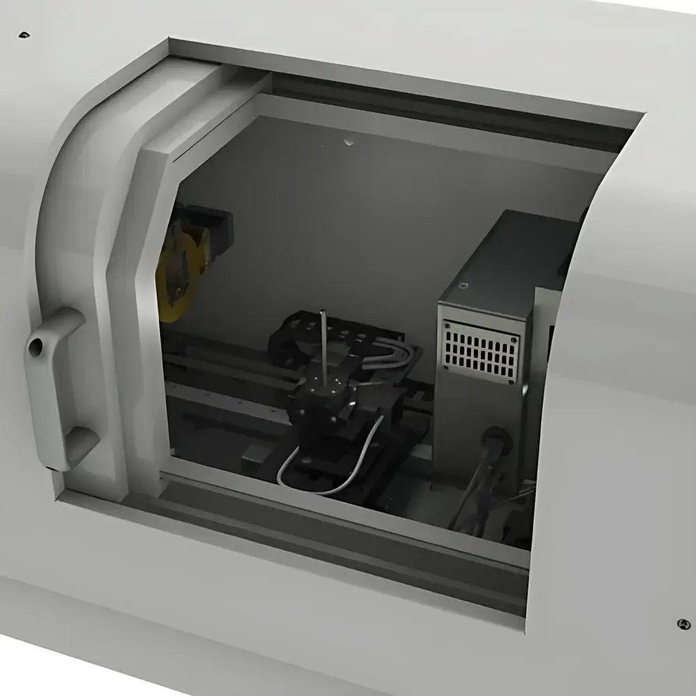
| Detector Type | High-Resolution CMOS-Based Direct-Conversion Semiconductor Detector (BrillianSe™) |
| Scan Mode | Rotation-Only (RO) |
| Spatial Resolution | Up to 5.6 µm (0.1 MTF at 90 cycles/mm) |
| X-ray Source Spot Size | ≤5 µm |
| X-ray Energy Range | 40–100 kV |
| Maximum Sample Volume | Ø25 mm × 100 mm (diameter × height/length) |
| System Dimensions | 1500 mm × 580 mm × 500 mm (L × W × H) |
| Pixel Pitch | 8 µm |
| Detector Format | 16 MP (4096 × 4096) |
| Field of View | 32 mm × 32 mm |
| Phase-Contrast Mechanism | Propagation-Based (Grating-Free, In-Line) |
Overview
The KA Imaging inCiTe Desktop Phase-Contrast MicroCT System is a high-resolution, benchtop 3D X-ray microscope engineered for quantitative non-destructive evaluation of low-absorbing, low-Z materials—such as biological tissues, polymers, composites, and additive-manufactured components. Unlike conventional absorption-based micro-CT systems, the inCiTe leverages propagation-based phase-contrast imaging (PCI), a coherent X-ray technique that converts subtle phase shifts induced by sample refractive index gradients into detectable intensity variations at the detector plane. This physical principle enables orders-of-magnitude improvement in contrast sensitivity for weakly attenuating specimens where traditional absorption contrast yields insufficient signal-to-noise ratio. At its core resides the BrillianSe™ direct-conversion detector—a proprietary amorphous selenium (a-Se)/CMOS hybrid sensor developed by KA Imaging. Its 8 µm pixel pitch, 16 MP resolution, and high detective quantum efficiency (DQE) across 40–120 keV allow high-fidelity, grating-free phase contrast imaging without optical coupling losses or light spread inherent in scintillator-based indirect detectors. The system integrates a microfocus X-ray source with ≤5 µm focal spot size, enabling true sub-6 µm spatial resolution (validated at 0.1 MTF, 90 cycles/mm) under optimized geometric magnification and acquisition conditions.
Key Features
- Propagation-based (in-line) phase-contrast imaging—no gratings, no alignment complexity, no flux penalty
- BrillianSe™ direct-conversion detector: 4096 × 4096 pixels, 8 µm pitch, high DQE up to 120 keV
- Microfocus X-ray source with ≤5 µm spot size for optimal geometric magnification and resolution scalability
- Rotation-only (RO) scanning geometry—mechanically robust, vibration-minimized, ideal for long-term stability
- Benchtop footprint (1500 × 580 × 500 mm) with full shielding compliant with IEC 61331-1 and local radiation safety regulations
- Native support for both absorption and phase-contrast reconstruction workflows within a single acquisition session
- No sample preparation required: no staining, sectioning, or contrast agents needed for soft-tissue or polymer imaging
Sample Compatibility & Compliance
The inCiTe accommodates cylindrical samples up to Ø25 mm × 100 mm in height—compatible with standard NDT test pieces, preclinical animal models (e.g., murine knee joints), pharmaceutical capsules, electronic assemblies, geological cores, and additively manufactured metal or polymer parts. Its phase-sensitive detection mechanism delivers superior contrast for materials with low atomic number (Z) and minimal X-ray attenuation—such as hydrogels, collagen matrices, carbon-fiber composites, aramid fabrics, and lightweight concrete aggregates. The system meets ISO 17025 requirements for measurement traceability when operated with calibrated phantoms (e.g., Gammex CT Performance Phantom). Data acquisition and reconstruction workflows support audit-ready metadata logging aligned with GLP and GMP documentation practices. While not FDA-cleared for clinical use, the inCiTe complies with IEC 62464-1 (medical X-ray equipment standards) and supports 21 CFR Part 11-compliant software modules for electronic signatures and audit trails when deployed in regulated R&D environments.
Software & Data Management
Acquisition and reconstruction are managed through KA Imaging’s inCiTe Control Suite—a Qt-based, multi-threaded application with intuitive GUI designed for both novice operators and advanced users. System boot-up to first scan takes under 20 minutes; automatic beam alignment, flat-field correction, and center-of-rotation calibration are performed in <60 seconds. Projection data are acquired in DICOM 3.0 format and stored with embedded metadata (kV, µA, exposure time, rotation step, detector gain, temperature). Reconstruction leverages GPU-accelerated filtered back-projection (FBP) and iterative algorithms (SART, OS-SART) supporting ring artifact suppression, beam-hardening correction, and phase-retrieval filtering (Paganin method). Output volumes are exported as 16-bit TIFF stacks or NIfTI files, fully compatible with commercial segmentation tools (Avizo, Dragonfly, Amira) and open-source platforms (3D Slicer, Fiji/ImageJ). All user actions—including parameter changes, reconstructions, and export events—are logged with timestamps and operator IDs for full traceability.
Applications
- Microstructural characterization of porous media, foams, and fiber-reinforced composites
- Non-destructive metrology for GD&T validation and reverse engineering of legacy parts
- Porosity analysis and defect mapping in laser-powder bed fusion (LPBF) and binder jetting components
- Clinical preclinical imaging: cartilage degeneration, vascular calcification, renal stone morphology
- Pharmaceutical solid-dose analysis: capsule coating uniformity, tablet internal cracking, excipient distribution
- Electronics inspection: solder joint integrity, wire bond voids, PCB delamination
- Geological core analysis: pore network topology, fluid saturation dynamics, mineral phase segmentation
FAQ
Does the inCiTe require synchrotron radiation to perform phase-contrast imaging?
No. The inCiTe achieves high-sensitivity phase contrast using laboratory-scale microfocus X-ray sources and propagation-based geometry—no synchrotron, no crystal optics, no interferometric setup required.
Can the system reconstruct both absorption and phase-contrast volumes from the same dataset?
Yes. Dual-contrast reconstruction is supported via post-processing: absorption contrast uses standard FBP, while phase contrast applies Paganin-type phase retrieval prior to reconstruction.
What is the minimum detectable feature size under typical operating conditions?
The system achieves 5.6 µm spatial resolution (0.1 MTF at 90 cycles/mm) with a 5 µm focal spot source, 1× geometric magnification, and optimal detector binning—verified using line-pair phantoms per ASTM E2737.
Is the BrillianSe™ detector suitable for high-energy applications beyond 100 kV?
Yes. BrillianSe™ maintains high DQE up to 120 keV, enabling extended energy range usage for dense composite inspection or thick-section imaging—subject to appropriate filtration and dose optimization.
How does the inCiTe handle beam hardening artifacts in polychromatic spectra?
The software includes dual-energy pre-correction models and iterative reconstruction with spectrum-aware forward projection, reducing cupping and streaking in heterogeneous samples.


")