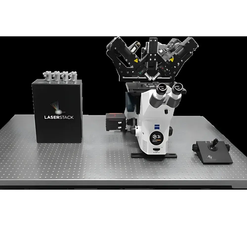
3i Marianas LightSheet DiSPIM Microscope
| Brand | 3i |
|---|---|
| Origin | USA |
| Model | Marianas LightSheet |
| Imaging Principle | Dual-Orthogonal Selective Plane Illumination Microscopy (diSPIM) |
| Spatial Resolution | 330 nm isotropic (x/y/z) |
| Maximum Frame Rate | 600 fps |
| Environmental Control | Enclosed incubation chamber (15–40 °C, CO₂, humidity) |
| Software Platform | SlideBook 6.0 |
| Compatible Sample Formats | Standard glass-bottom dishes, coverslips, multi-well plates |
| Excitation Lasers (Standard) | 405 nm, 488 nm, 593 nm, 640 nm |
| Modular Expansion Options | Spinning-disk confocal, TIRF, FLIM, optogenetic stimulation units |
Overview
The 3i Marianas LightSheet (MLS) is a high-performance, inverted light sheet fluorescence microscope engineered for long-term, high-fidelity 3D dynamic imaging of living specimens. Built upon dual-orthogonal selective plane illumination microscopy (diSPIM), the system delivers true isotropic resolution by intersecting two orthogonally oriented light sheets—each generated and detected through independent 45°-tilted objectives—followed by computationally fused deconvolution. Unlike conventional single-axis light sheet systems, diSPIM eliminates axial resolution asymmetry and mitigates scattering-induced artifacts, enabling quantitative structural reconstruction of subcellular features across all three dimensions. The MLS is purpose-built for live-cell applications where phototoxicity, temporal fidelity, and physiological relevance are non-negotiable constraints. Its optical architecture minimizes out-of-plane excitation, reducing photon dosage by >95% compared to confocal or widefield fluorescence microscopy—thereby preserving native cell behavior over hours to days without compromising signal-to-noise ratio.
Key Features
- diSPIM dual-view acquisition: Two synchronized, independently controlled objective paths provide orthogonal illumination and detection. Real-time image fusion and iterative deconvolution yield isotropic 330 nm resolution in x, y, and z—enabling accurate volumetric quantification of organelle dynamics, cytoskeletal remodeling, and intercellular interfaces.
- High-speed volumetric imaging: Piezo-driven objective scanning enables rapid light sheet translation through the specimen. Combined with sCMOS camera readout optimization, the system achieves up to 600 frames per second at full field-of-view—sufficient to resolve millisecond-scale events including vesicle trafficking, calcium wave propagation, and mitotic spindle assembly.
- Integrated environmental control: A fully enclosed, temperature-, CO₂-, and humidity-regulated incubation chamber maintains physiological conditions throughout extended acquisitions. Active feedback loops ensure stability within ±0.2 °C and ±0.1% CO₂—validated for uninterrupted imaging of sensitive primary cultures and developing embryos.
- Multi-modal modularity: The platform natively supports widefield fluorescence, brightfield, and DIC imaging. Optional modules—including spinning-disk confocal, TIRF, time-resolved FLIM, and patterned optogenetic stimulation—can be added without hardware realignment or software reconfiguration.
- Universal sample compatibility: Horizontal sample loading accommodates standard glass-bottom dishes (e.g., MatTek, Ibidi), coverslips, and multi-well plates. No capillary embedding, tissue clearing, or specialized mounting is required—enabling direct transition from routine cell culture to 3D time-lapse imaging.
- Multi-wavelength laser architecture: Four solid-state lasers (405, 488, 593, 640 nm) are pre-aligned and intensity-stabilized. Additional wavelengths (e.g., 561 nm, 730 nm) can be integrated via fiber-coupled ports while maintaining beam collimation and power consistency across modalities.
Sample Compatibility & Compliance
The Marianas LightSheet accepts specimens ranging from single adherent cells and 3D organoids to zebrafish and Drosophila embryos (up to 1.5 mm in diameter). Its open-access stage design permits use with commercially available microfluidic chambers, perfusion systems, and electrophysiology rigs. All optical components comply with ISO 10110-7 surface quality standards; laser safety meets IEC 60825-1 Class 1 requirements when interlocked. The system supports GLP/GMP-aligned workflows: SlideBook 6.0 implements audit-trail logging, electronic signatures, and user-access controls compliant with FDA 21 CFR Part 11 for regulated environments.
Software & Data Management
SlideBook 6.0 serves as the unified control and analysis environment. It orchestrates hardware synchronization (lasers, shutters, piezo stages, cameras), acquires multi-channel, multi-timepoint datasets, and applies GPU-accelerated deconvolution using constrained iterative algorithms. Built-in tools include skew correction for oblique imaging geometries, automatic drift compensation, 3D surface rendering (isosurface, volume rendering), and quantitative colocalization metrics (Manders’ coefficients, Pearson correlation). Raw data is saved in TIFF or OME-TIFF format with embedded metadata (acquisition parameters, calibration files, stage positions), ensuring FAIR (Findable, Accessible, Interoperable, Reusable) data principles. Batch processing pipelines support parallelized analysis across hundreds of time-lapse stacks.
Applications
- Cell biology: Long-term tracking of mitosis, migration, autophagy, and endosomal trafficking in unperturbed monolayers or spheroids; quantification of nuclear envelope rupture, mitochondrial fission/fusion cycles, and ER morphology.
- Developmental biology: Whole-embryo time-lapse imaging of gastrulation, neurulation, and organogenesis in zebrafish and Drosophila—with minimal photodamage and no need for embryo immobilization or chemical anesthetics.
- Neuroscience: Simultaneous optogenetic stimulation and functional imaging in cultured neurons or cleared brain slices; mapping of dendritic spine turnover, axonal transport kinetics, and synaptic vesicle recycling.
- Drug discovery: Phenotypic screening of compound libraries on live 3D tumor spheroids or iPSC-derived cardiomyocytes; kinetic assessment of cytotoxicity, metabolic perturbation, and pathway activation using ratiometric biosensors.
- Plant and microbiology: Imaging of root hair development, pollen tube growth, and bacterial biofilm architecture under near-native hydration and gas exchange conditions.
FAQ
Is the Marianas LightSheet compatible with existing inverted microscopes?
Yes—the system is designed as a modular upgrade path. Its optical train and stage interface can be retrofitted onto select Nikon Eclipse Ti and Zeiss Axio Observer platforms using OEM-certified adapter kits.
Does SlideBook support batch processing of large diSPIM datasets?
Yes—SlideBook 6.0 includes distributed computing support via local clusters or cloud-based HPC resources. Deconvolution, registration, and segmentation workflows can be scripted in Python or executed through the GUI with parameter inheritance across experiments.
Can the system perform multi-position time-lapse across a 96-well plate?
Yes—integrated motorized XY stage and autofocus routines enable automated multi-site acquisition with drift correction between wells. Plate maps are imported directly from LIMS or Excel.
What is the minimum recommended sample thickness for optimal diSPIM performance?
Optimal performance is achieved for samples 20–300 µm thick. Thicker specimens (>500 µm) benefit from optional adaptive optics correction or refractive-index-matched mounting media.
Are service contracts and application support included with purchase?
3i provides comprehensive installation, validation, and on-site training. Extended service agreements include remote diagnostics, priority parts dispatch, and dedicated application scientist consultation for experimental design and data interpretation.


