
Biological Microscope/In Vivo Imaging
Filter
Showing 271–300 of 424 results
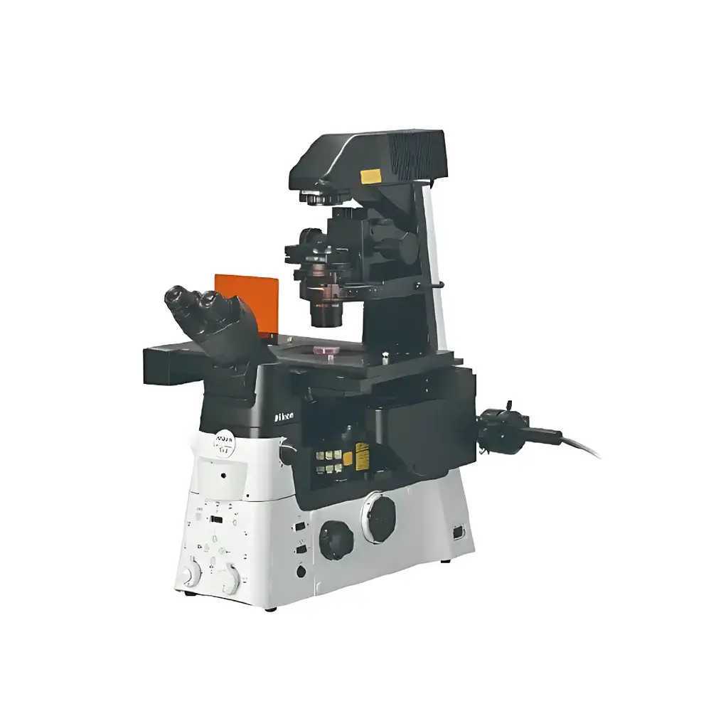
| Brand | Nikon |
|---|---|
| Origin | Japan |
| Model | ECLIPSE Ts2 |
| Optical System | CFI60 Infinity-Corrected |
| Observation Modes | Brightfield, Phase Contrast (including Plan Apochromat Phase), DIC-like Relief Contrast*¹ |
| Fluorescence Capability | Yes (Ts2-FL variant) |
| Illumination | High-Brightness White LED Transmitted Light (with Köhler illumination via built-in fly-eye lens) |
| Eyepiece Angle | 45°, Interpupillary Distance: 50–75 mm (Siedentopf), Port Split: 100/0 or 0/100 |
| Focusing Mechanism | Objective Nosepiece Movement (7 mm up / 1.5 mm down), Coarse Focus: 37.7 mm/rev, Fine Focus: 0.2 mm/rev, Adjustable Coarse Focus Torque |
| Objective Turret | 5-Position |
| Condenser | ELWD (NA 0.3, WD 75 mm) |
| Stage | Flat Stage (170 × 247 mm) |
| Dimensions (W×D×H) | 236 × 548 × 471 mm (Ts2), 236 × 564 × 471 mm (Ts2-FL) |
| Weight | ~13 kg (Ts2), ~14.5 kg (Ts2-FL) |
| Power Supply | 100–240 V AC ±10%, 50/60 Hz, 0.35 A |
| Power Consumption | 15 W |
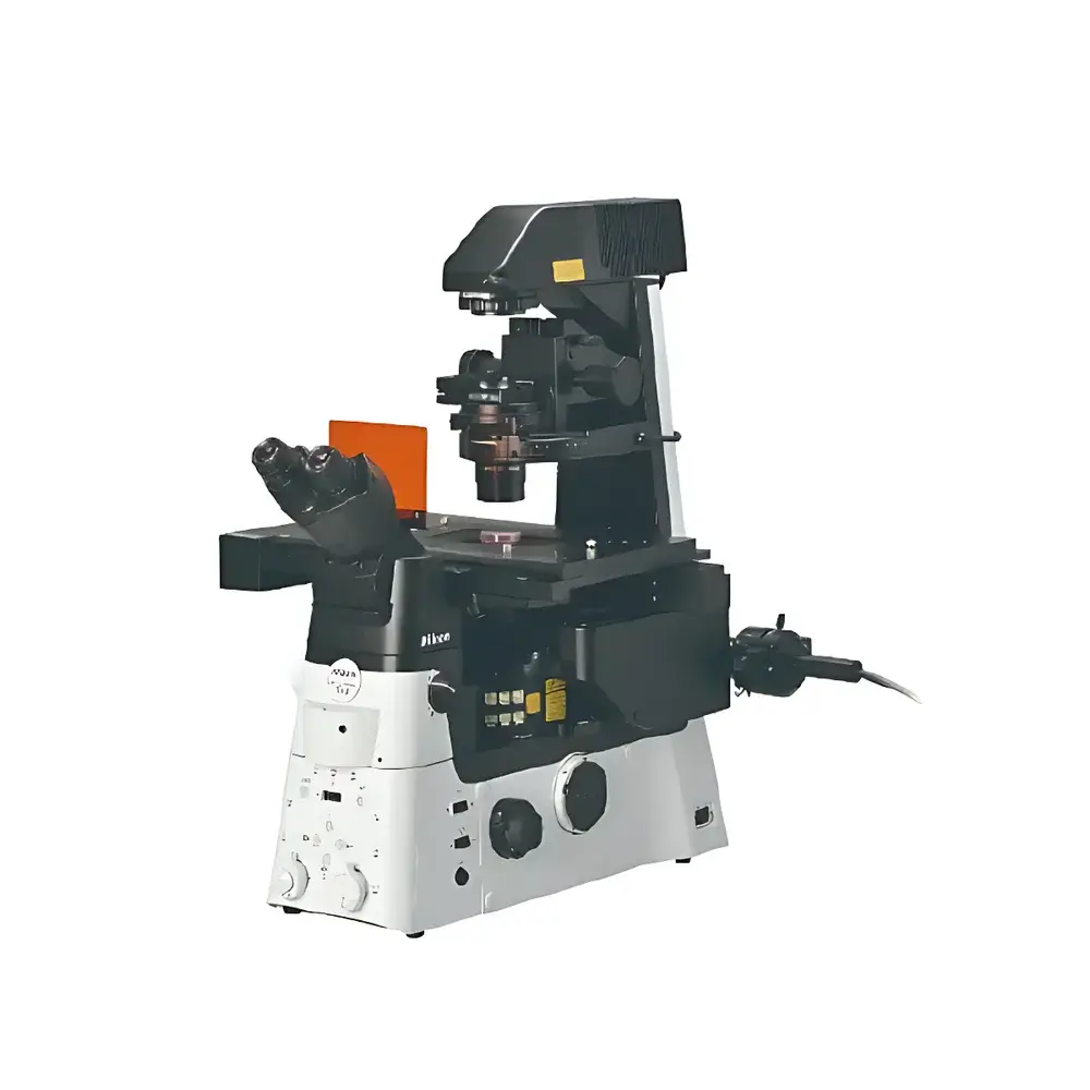
| Brand | Nikon |
|---|---|
| Origin | Japan |
| Model | ECLIPSE Ts2-FL |
| Optical System | CFI60 Infinity-Corrected |
| Observation Modes | Brightfield, Phase Contrast (T-PH), Differential Interference Contrast (DIC)-like Relief Contrast, Epifluorescence |
| Illumination | High-Brightness White LED Transmitted Light + Modular LED Epifluorescence (385, 455, 470, 505, 525, 560, 590, 625 nm) |
| Eyepiece Field Number | 22 (10×), 16 (15×), 12.5 (20×) |
| Tube Angle | 45°, Interpupillary Distance: 50–75 mm (Siedentopf), Trinocular Port (100/0 or 0/100 beam splitter) |
| Focus Travel | 8.5 mm total (7 mm upward, 1.5 mm downward) |
| Coarse Focus | 37.7 mm/rev, Fine Focus: 0.2 mm/rev, Adjustable Coarse Focus Torque |
| Objective Turret | 5-Position |
| Condenser | ELWD (NA 0.3, WD 75 mm) with Pre-centered and Centerable Phase Contrast Sliders (for 10×, 20×, 40×) and Relief Contrast Sliders (dual-side configuration required) |
| Stage | Fixed Plate Stage (170 × 247 mm) with Acrylic Circular Cover Slip |
| Compatible with Multiple Sample Holders | CS-HP35 (35 mm dish), CS-HLP100 (100 mm dish), CS-HT (Terasaki plate & Ø65 mm dish), CS-HS (slide, Ø54 mm dish, hemocytometer), C-S-HU (universal holder for Terasaki, slides, Ø35–65 mm dishes, hemocytometer), C-S-HG (circular glass slide holder), C-S-HLS (circular cover slip kit) |
| Fluorescence Attachment | Motorized Filter Cube Turret (3 positions + brightfield slot), Integrated Noise-Reduction Mechanism, Optional Light Shield |
| Dimensions (W×D×H) | 236 × 548 × 471 mm (Ts2), 236 × 564 × 471 mm (Ts2-FL) |
| Weight | ~13 kg (Ts2), ~14.5 kg (Ts2-FL) |
| Power Supply | 100–240 VAC ±10%, 50/60 Hz, 0.35 A |
| Power Consumption | 15 W |
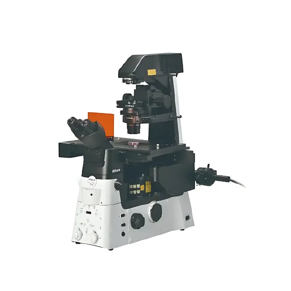
| Brand | Nikon |
|---|---|
| Origin | Japan |
| Model | ECLIPSE Ts2R |
| Optical System | CFI60 Infinity-Corrected |
| Observation Modes | Brightfield, Phase Contrast (including TMC), DIC, NAMC (Nikon Advanced Modulation Contrast), Relief Contrast, Fluorescence Epi-Illumination, Spindle Observation |
| Illumination | High-Brightness White LED Transmitted Light (with Homogenizing Lens), LED Epi-Illumination (up to 4 interchangeable units |
| wavelengths | 385, 455, 470, 505, 525, 590, 625 nm) |
| Eyepiece Options | 10× (22 mm FOV), 12.5× (16 mm), 15× (14.5 mm) |
| Binocular Tube | Siedentopf-type, interpupillary distance 50–75 mm, tilt angle 35° (standard) or 15–45° (ergonomic) |
| Camera Port Split Ratios | 100:0 / 0:100, 100:0 / 20:80, 100:0 / 50:50 |
| Focus Mechanism | Objective nosepiece-driven Z-movement |
| coarse focus | 5.0 mm/rev, fine focus: 0.1 mm/rev |
| Objective Turret | 6-position, with DIC prism slots |
| Condenser | Rotating condenser turret (7-slot), compatible with ELWD, LWD, and NAMC condensers |
| Stage | Flat stage (260 × 300 mm) |
| Stage Adapters | C-S-HP35 (35 mm dish), C-S-HLP100 (100 mm dish), C-S-HT (Terasaki plates & Ø65 mm dishes), C-S-HS (slide, Ø54 mm dish, hemocytometer), C-S-HU (universal for Terasaki, slides, Ø35–65 mm dishes, hemocytometer), C-S-HG (circular glass slide), C-S-HLS (circular slide kit), TC-S-HA (acrylic stage plate) |
| Fluorescence Accessories | Motorized filter cube turret (body-integrated, noise-reduction design) |
| Dimensions (W×D×H) | 286 × 466 × 542 mm |
| Weight | ~17–18 kg |
| Power Supply | 100–240 V AC, <0.65 A |
| Power Consumption | 30 W |
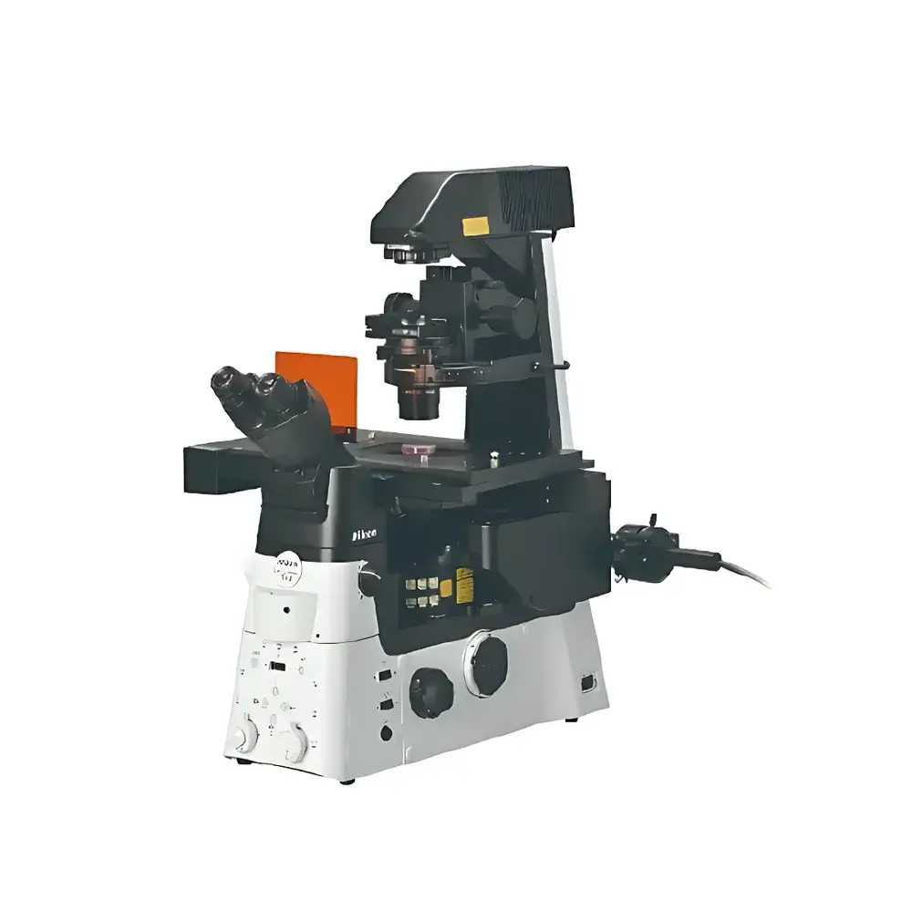
| Brand | Nikon |
|---|---|
| Origin | Japan |
| Model | ECLIPSE Ts2R-FL |
| Optical System | CFI60 Infinity-Corrected |
| Observation Modes | Brightfield, Phase Contrast (including TIRF-capable phase), DIC, NAMC (Nikon Advanced Modulation Contrast), Relief Contrast, Spindle Observation, Epifluorescence |
| Illumination | High-Intensity White LED Transmitted Light (with Köhler illumination and built-in fly-eye lens) |
| wavelengths | 385, 455, 470, 505, 525, 590, 625 nm) |
| Eyepiece Options | 10×/22, 12.5×/16, 15×/14.5 field number |
| Trinocular Port Split Ratios | 100:0 / 0:100, 100:0 / 20:80, 100:0 / 50:50 |
| Focus Mechanism | Objective nosepiece-driven Z-axis movement (8 mm upward, 3 mm downward travel) |
| coarse focus | 5.0 mm/rev, fine focus: 0.1 mm/rev |
| Nosepiece | 6-position revolver with DIC prism slots |
| Condenser | Multi-position condenser turret (7-slot), compatible with ELWD, LWD, and NAMC condensers |
| Stage | Flat stage (260 × 300 mm) |
| Fluorescence Accessories | Motorized filter cube turret (with noise-reduction mechanism) |
| Dimensions (W×D×H) | 286 × 466 × 542 mm |
| Weight | ~17–18 kg |
| Power Supply | 100–240 V AC, <0.65 A, 30 W max |
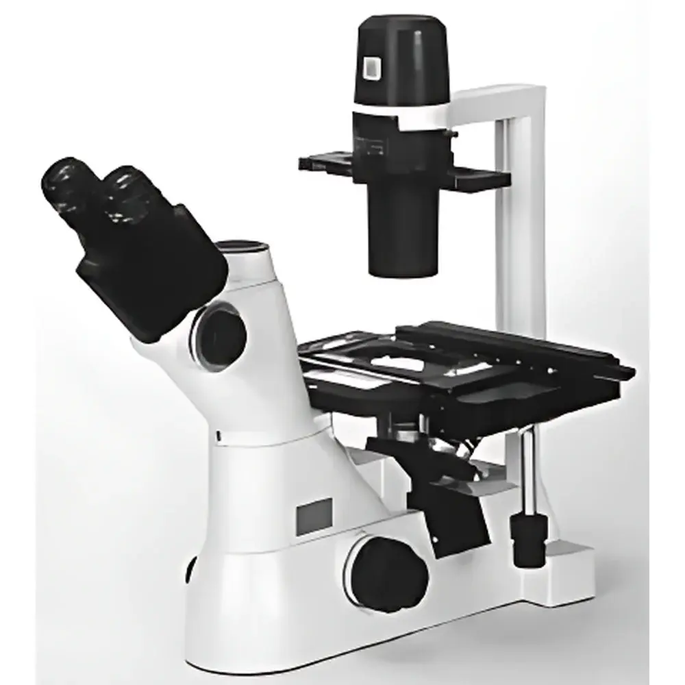
| Brand | Nikon |
|---|---|
| Origin | Germany |
| Model | TS2 |
| Microscope Type | Inverted Microscope |
| Eyepiece Configuration | Trinocular |
| Optical System | CFI60 Infinity-Corrected |
| Focusing Mechanism | Coaxial Coarse/Fine Adjustment |
| Working Distance | Up to 75 mm (with LWD condenser) |
| Condenser Options | Long Working Distance (NA 0.3, WD 75 mm) and Hoffman Modulation Contrast (NA 0.4, WD 44 mm) |
| Fluorescence Capability | Optional Epi-Fluorescence Attachment (up to two filter cubes, 50 W mercury lamp) |
| Camera Port | Standard C-mount compatible port for Nikon digital cameras and third-party imaging systems |
| Ergonomic Design | 45° inclined trinocular head, 400 mm eye-point height, adjustable interpupillary distance (50–75 mm) |
| Stage Height | 195 mm above bench surface |
| Stage Clearance | 190 mm between stage and condenser |
| Phase Contrast | Equipped with Nikon ADL (Apodized Diffraction-Limited) Phase Contrast Objectives |
| Compliance | Designed for GLP-compliant documentation workflows |
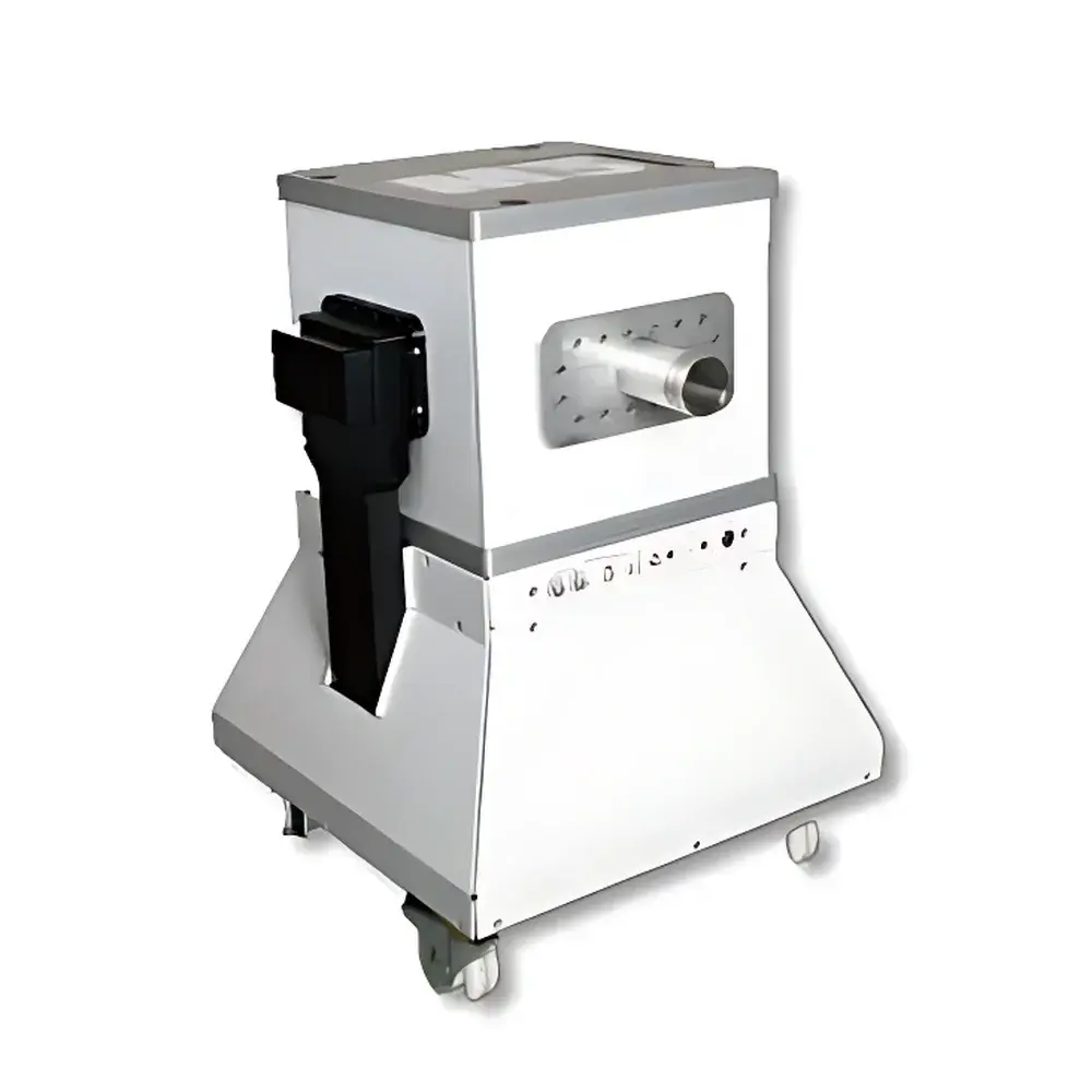
| Brand | NIUMAG |
|---|---|
| Origin | Israel |
| Manufacturer Type | Authorized Distributor |
| Origin Category | Imported |
| Model | M3 |
| Instrument Type | Preclinical Magnetic Resonance Imaging (MRI) System |
| Magnet Type | Permanent Magnet, 1.0 T |
| Spatial Resolution | ≤ 235 µm (isotropic) |
| Scan Time | As low as 8–9 min per sequence |
| Cooling | Passive (No Cryogens or Chiller Required) |
| Shielding | Self-Shielded, Zero Stray Field |
| Regulatory Compliance | CE Marked, Designed for GLP-Compliant Preclinical Research |
| Application Focus | Mouse Phenotyping, Longitudinal In Vivo Imaging |
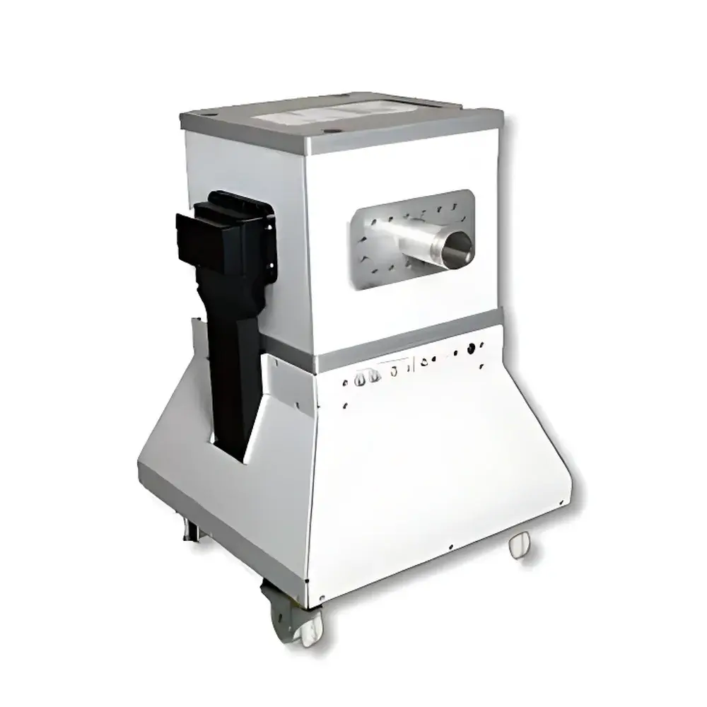
| Brand | NIUMAG |
|---|---|
| Origin | Israel |
| Manufacturer Type | Authorized Distributor |
| Origin Category | Imported |
| Model | M3 |
| Instrument Type | Nuclear Magnetic Resonance (NMR) Imaging System |
| Magnet Type | Permanent Magnet, 1.0 Tesla |
| Field Homogeneity | <10 ppm over 20 mm DSV |
| Gradient Strength | ≥200 mT/m |
| Gradient Slew Rate | ≥1000 T/m/s |
| Spatial Resolution | ≤200 µm isotropic (typical in vivo mouse brain) |
| Scan Time | <10 minutes per standard sequence |
| Cooling | Passive (no cryogens or chiller required) |
| Shielding | Self-shielded, zero fringe field beyond instrument footprint |
| Regulatory Compliance | CE Marked, IEC 61000-6-3/6-4, ISO 13485–certified manufacturing processes |
| Software Platform | Aspect ImageSuite v5.x (FDA 21 CFR Part 11 compliant audit trail optional) |
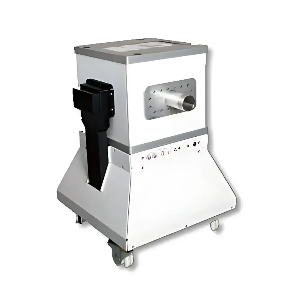
| Brand | NIUMAG |
|---|---|
| Origin | Israel |
| Manufacturer Type | Authorized Distributor |
| Origin Category | Imported |
| Model | M3-mice |
| Instrument Type | Preclinical Magnetic Resonance Imaging (MRI) System |
| Magnet Type | Permanent Magnet, 1.0 Tesla |
| Spatial Resolution | ≤ 235 µm (isotropic, typical in vivo) |
| Scan Time | As low as 8–9 minutes per sequence (T1- or T2-weighted) |
| Cooling | Passive (no cryogens or chillers required) |
| Shielding | Self-shielded, zero fringe field beyond system footprint |
| Compliance | Designed for ISO 13485-aligned manufacturing and GLP-compliant preclinical research environments |
| Software Platform | Aspect ImageSuite™ v5.x (FDA 21 CFR Part 11–ready audit trail, DICOM 3.0 export, multi-sequence reconstruction pipeline) |
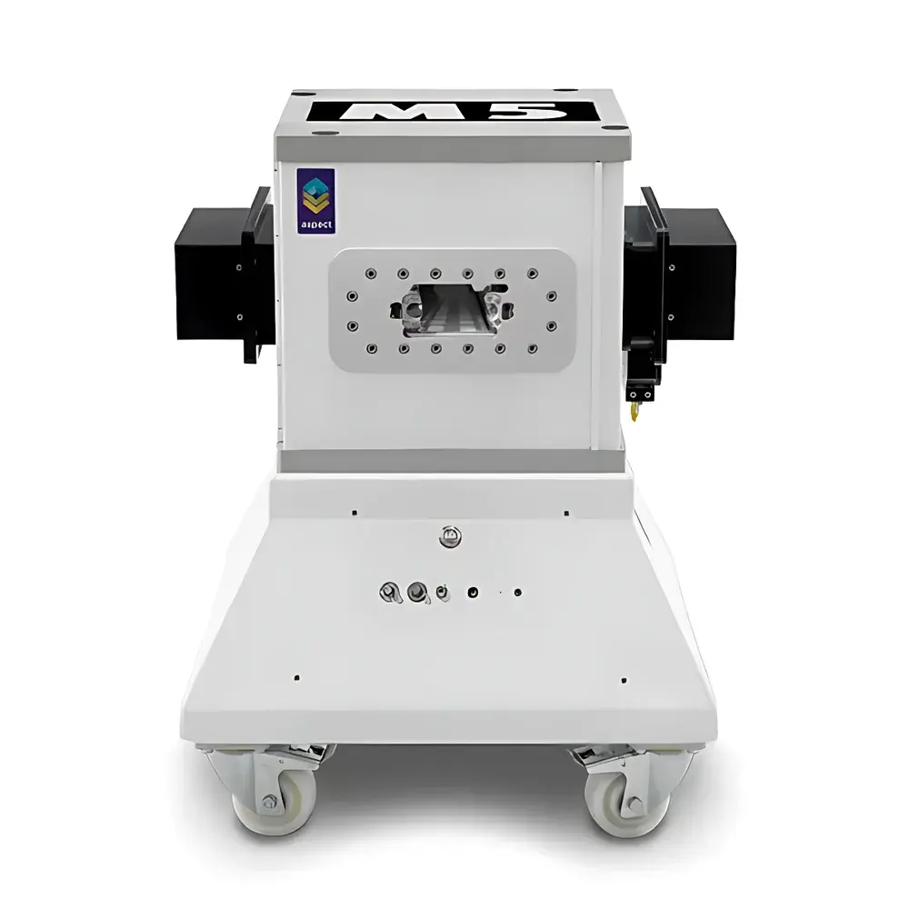
| Brand | NIUMAG |
|---|---|
| Origin | Israel |
| Manufacturer Type | Authorized Distributor |
| Origin Category | Imported |
| Model | M5 |
| Instrument Type | Nuclear Magnetic Resonance Imaging (MRI) System |
| Magnet Type | Permanent Magnet, 1 Tesla |
| Weight Capacity | Up to 700 g (mouse/rat) |
| Imaging Modes | 2D/3D, In Vivo, Ex Vivo, In Vitro |
| Cooling | Passive (No Cryogens or Chiller Required) |
| Shielding | Self-Shielded (Zero Stray Field) |
| Typical Resolution | ≤ 234 µm (T2-weighted), ≤ 195 µm (T1-weighted) |
| Acquisition Time | ~4.8–9.1 min per scan |
| Compliance | Designed for ISO 13485-aligned lab environments |
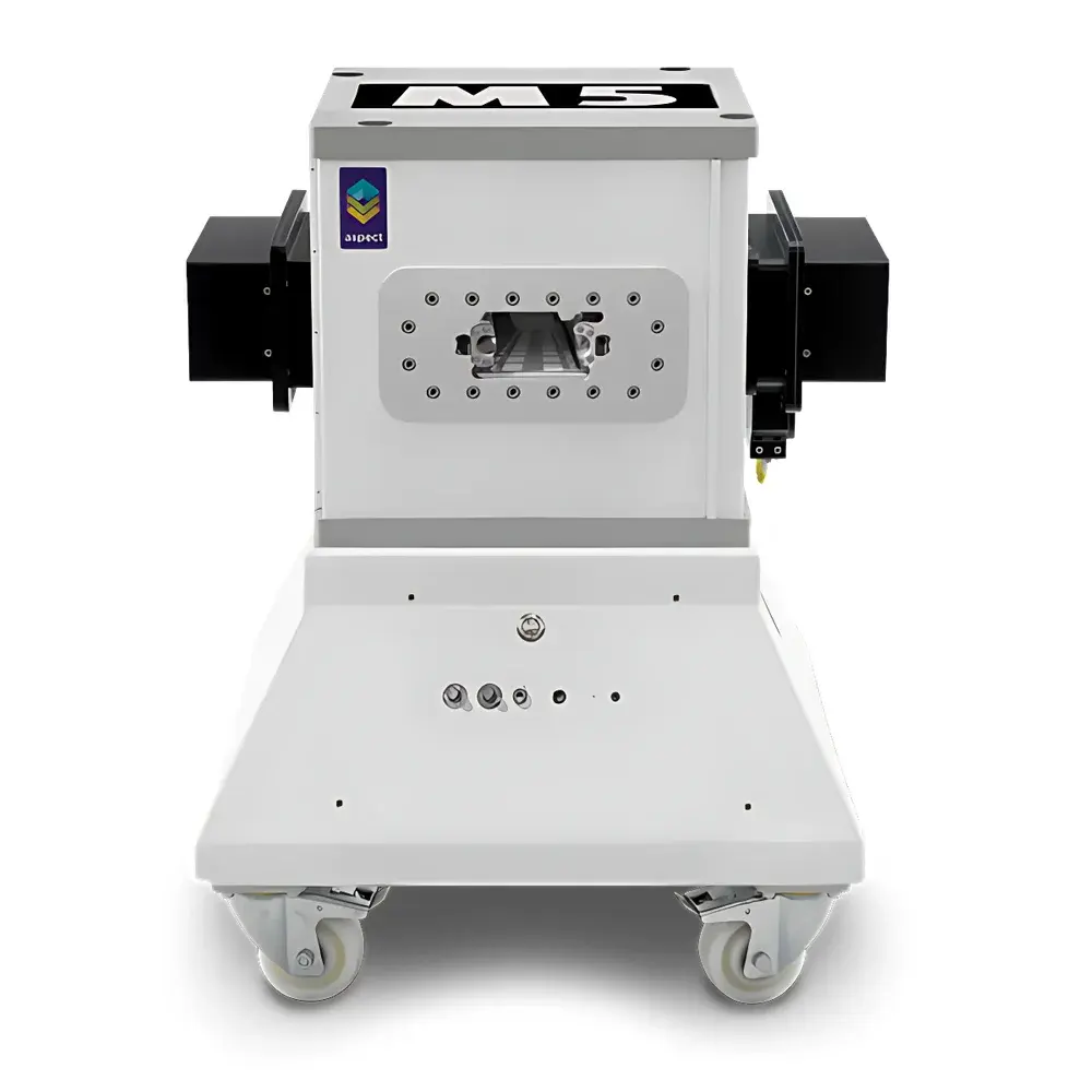
| Brand | NIUMAG |
|---|---|
| Origin | Israel |
| Manufacturer Type | Authorized Distributor |
| Origin Category | Imported |
| Model | M5-Rat |
| Instrument Type | Nuclear Magnetic Resonance (NMR) Imaging System |
| Magnet Type | Permanent Magnet, 1 Tesla |
| Spatial Resolution | ≤100 µm (in-plane, 3D gradient-echo mode) |
| Imaging Modes | T1-weighted, T2-weighted, PD-weighted, 2D/3D spin-echo and gradient-echo sequences |
| Application Focus | In vivo rodent musculoskeletal phenotyping, arthritis progression monitoring, preclinical OA model validation |
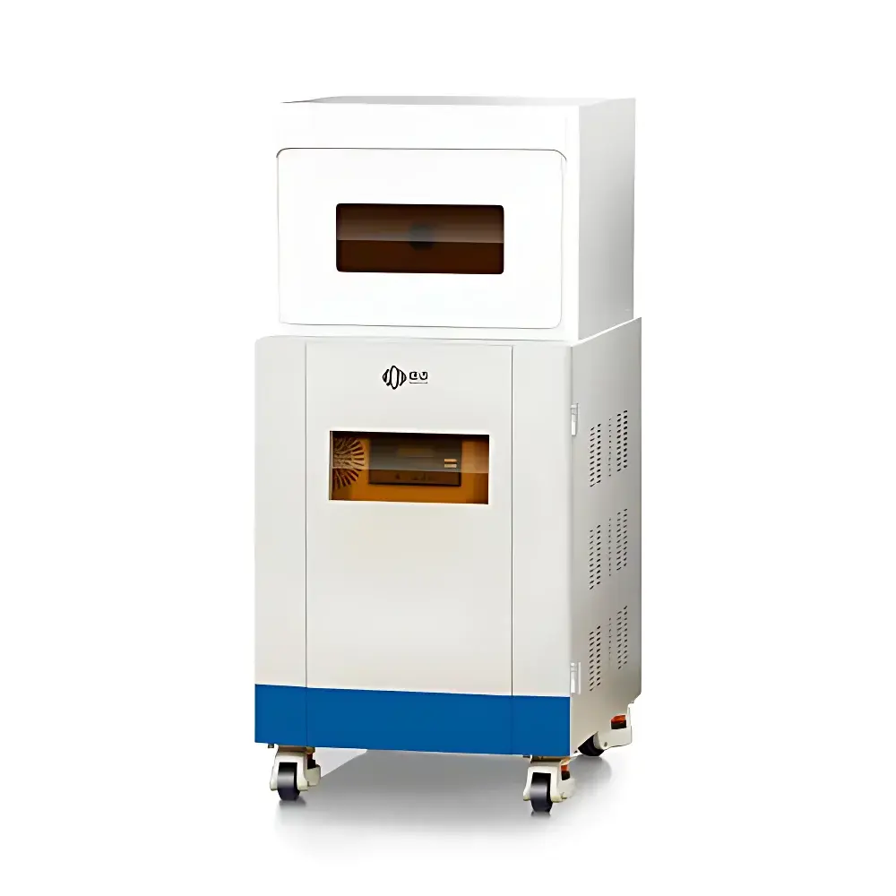
| Brand | NIUMAG |
|---|---|
| Origin | Shanghai, China |
| Manufacturer Type | Authorized Distributor |
| Region of Manufacture | Domestic (China) |
| Model | NM21-040H-I |
| Instrument Type | Benchtop Permanent-Magnet MRI System |
| Magnetic Field Strength | 0.5 ± 0.08 T |
| RF Coil Inner Diameter | 40 mm |
| Field of View (FOV) | 60 × 60 mm² |
| Maximum Sample Capacity | 1 animal per scan |
| Energy Resolution | Not applicable (N/A) |
| Spatial Resolution | Not specified in source |
| Scan Speed | Not quantified (system supports multi-sequence acquisition with variable TR/TE) |
| Compliance | Designed for preclinical GLP-compliant research environments |
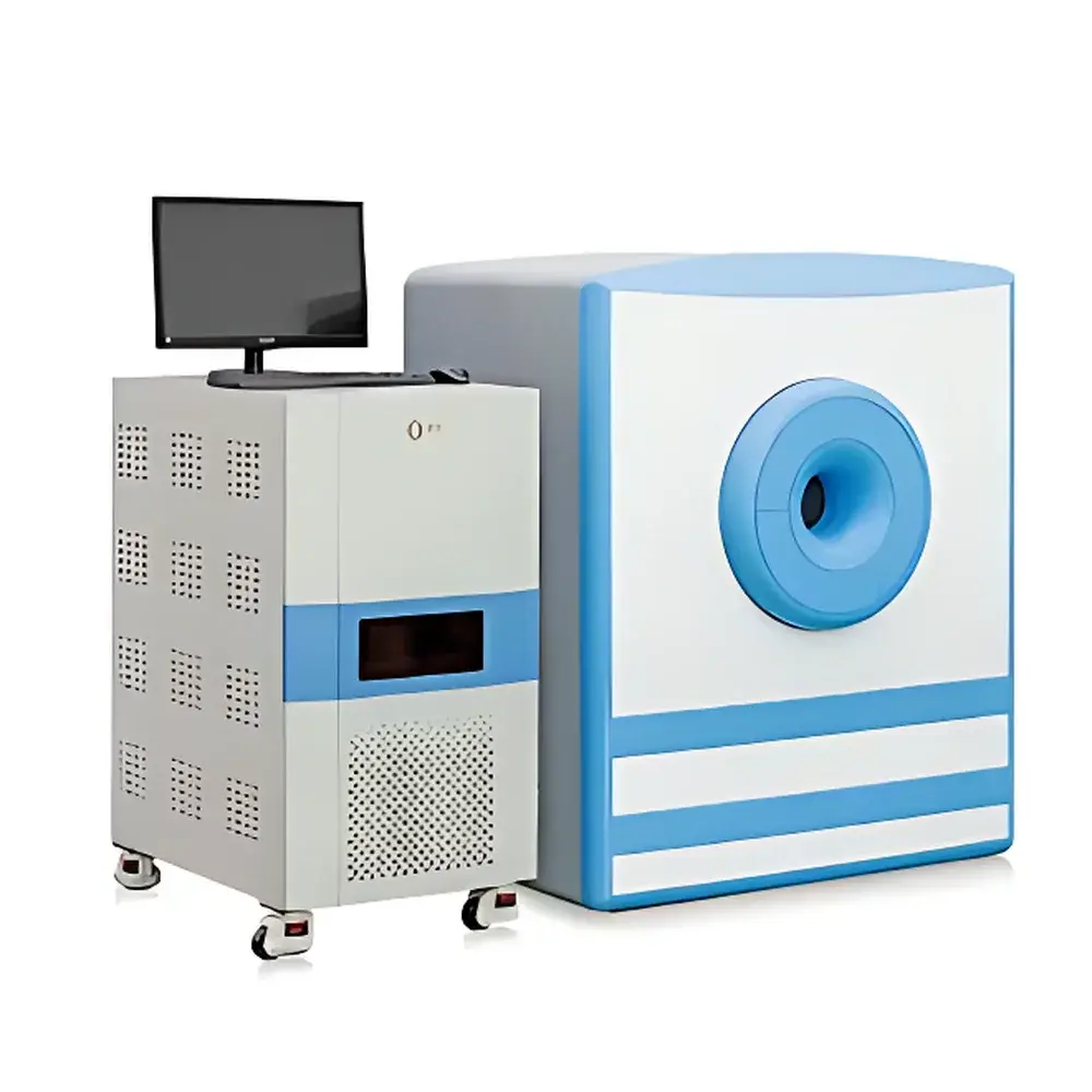
| Brand | NIUMAG |
|---|---|
| Origin | Jiangsu, China |
| Manufacturer Type | Authorized Distributor |
| Country of Origin | China |
| Model | NM42 |
| Instrument Type | 1.0 Tesla Permanent-Magnet Benchtop MRI System |
| Animal Weight Range | 1–45 g |
| Maximum Sample Width | < 40 mm |
| Minimum Sample Volume | ≥ 100 µL |
| Minimum Slice Thickness | 0.8 mm |
| Field Homogeneity | Optimized for high SNR and spatial resolution |
| Anesthesia Compatibility | Optional integrated gas anesthesia module |
| Shielding Requirement | None — no RF-shielded room required |
| Brand | NIUMAG |
|---|---|
| Origin | Jiangsu, China |
| Manufacturer Type | Authorized Distributor |
| Country of Origin | China |
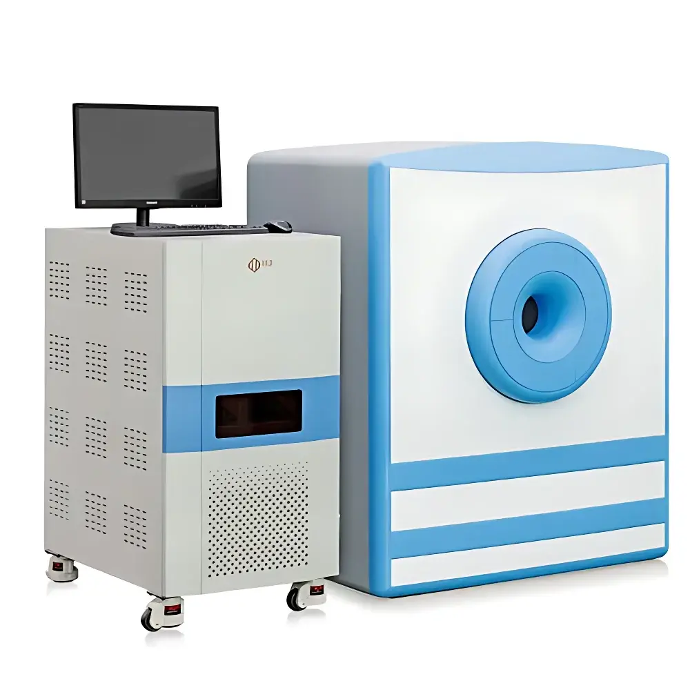
| Model | NM42-NIUMAG |
| Instrument Type | Permanent-Magnet Small Animal Magnetic Resonance Imaging System |
| Magnetic Field Strength | ~1.0 T (Permanent Magnet) |
| Bore Diameter | ≥40 mm |
| Maximum Sample Weight | 45 g |
| Minimum Sample Volume | ≥100 µL |
| Minimum Slice Thickness | 0.8 mm |
| Imaging Modes | T1-, T2-, PD-weighted, Gradient Echo, Spin Echo, Multi-slice 2D/3D acquisitions |
| Anesthesia Compatibility | Optional Integrated Gas Anesthesia System |
| Software Compliance | FDA 21 CFR Part 11–ready audit trail (optional configuration), GLP/GMP-compatible data logging |
| Brand | NIUMAG |
|---|---|
| Origin | Jiangsu, China |
| Manufacturer Type | Authorized Distributor |
| Country of Origin | China |
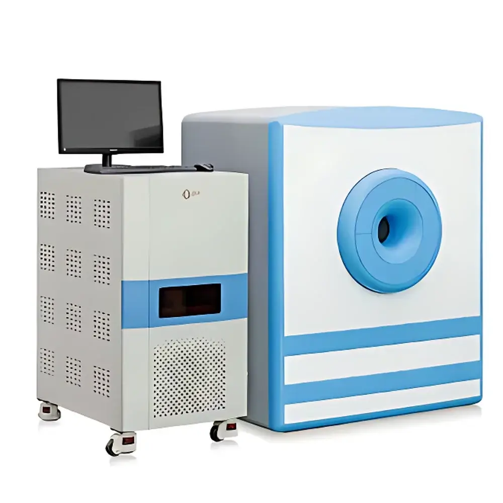
| Model | NM42- |
| Instrument Type | Benchtop Permanent-Magnet MRI System |
| Magnetic Field Strength | 1.0 T |
| Gradient Performance | Standard for Small Animal Imaging |
| Minimum Slice Thickness | 0.8 mm |
| Animal Weight Capacity | 1–45 g |
| Sample Chamber Diameter | ≤40 mm |
| Minimum Sample Volume | ≥100 µL |
| Imaging Modality | Proton (¹H) MRI |
| Safety | Non-ionizing, Non-invasive, No Radiation Exposure |
| Regulatory Compliance | Designed per ISO 13485-aligned manufacturing practices |
| Brand | NIUMAG |
|---|---|
| Origin | Jiangsu, China |
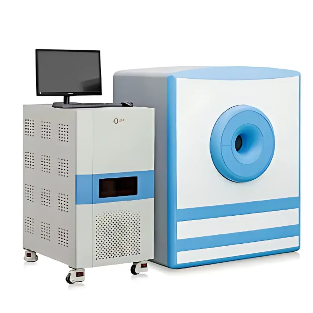
| Model | NM42-0 |
| Field Strength | 1.0 T Permanent Magnet |
| Maximum Sample Width | <40 mm |
| Animal Weight Range | 1–45 g |
| Minimum Sample Volume | ≥100 µL |
| Minimum Slice Thickness | 0.8 mm |
| Imaging Modality | Nuclear Magnetic Resonance (NMR)-based Magnetic Resonance Imaging (MRI) |
| Regulatory Classification | Preclinical In Vivo Imaging System |
| Compliance Framework | Designed for GLP-compliant preclinical research environments |
| Software | Integrated MRI acquisition platform with optional advanced image processing module |
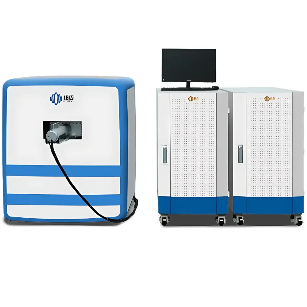
| Brand | NIUMAG |
|---|---|
| Origin | Shanghai, China |
| Manufacturer Type | Authorized Distributor |
| Country of Origin | China |
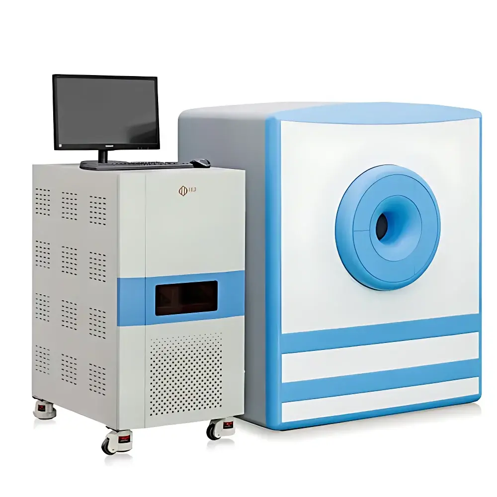
| Model | NM42-040H-I |
| Instrument Type | Magnetic Resonance Imaging (MRI) System |
| Magnetic Field Strength | 1.0 ± 0.08 T |
| Magnet Type | Permanent Magnet |
| Field Homogeneity | ≤20 ppm over Ø40 mm × H40 mm DSV |
| Imaging FOV | Ø40 mm × 40 mm (height) |
| Minimum Slice Thickness | 0.8 mm |
| Spatial Resolution (in-plane) | 0.08 mm |
| Gradient System | High-Performance Integrated Gradient Set |
| RF Coil Configuration | Animal-Specific Tunable RF Coils |
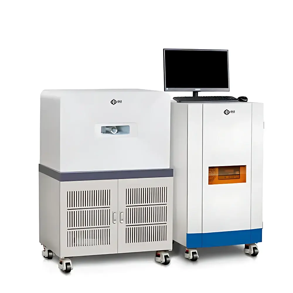
| Brand | NIUMAG |
|---|---|
| Origin | Jiangsu, China |
| Manufacturer Type | Authorized Distributor |
| Country of Origin | China |
| Model | NM42-060H-I-1 |
| Instrument Type | Low-Field Nuclear Magnetic Resonance (LF-NMR) Imaging System |
| Magnet Type | Permanent Magnet |
| Field Strength | 1.0 T ± 0.05 T |
| Homogeneity | ≤30 ppm over 60 mm DSV |
| Sample Capacity | 1–350 g (mice, rats, excised tissues) |
| Imaging Modes | T1-weighted, T2-weighted, Proton Density-weighted, Water-Fat Suppressed Imaging |
| Brand | NIUMAG |
|---|---|
| Origin | Jiangsu, China |
| Manufacturer Type | Authorized Distributor |
| Country of Origin | China |
| Model | QMR12-060H-I |
| Instrument Type | Low-Field Nuclear Magnetic Resonance (NMR) Analyzer |
| Spatial Resolution | 0.1 mm |
| Field of View (FOV) | 100 mm × 100 mm |
| Magnet Type | Permanent Magnet |
| Probe | Dedicated Body Composition RF Coil |
| Measurement Mode | Non-invasive, Anesthesia-Free, In Vivo |
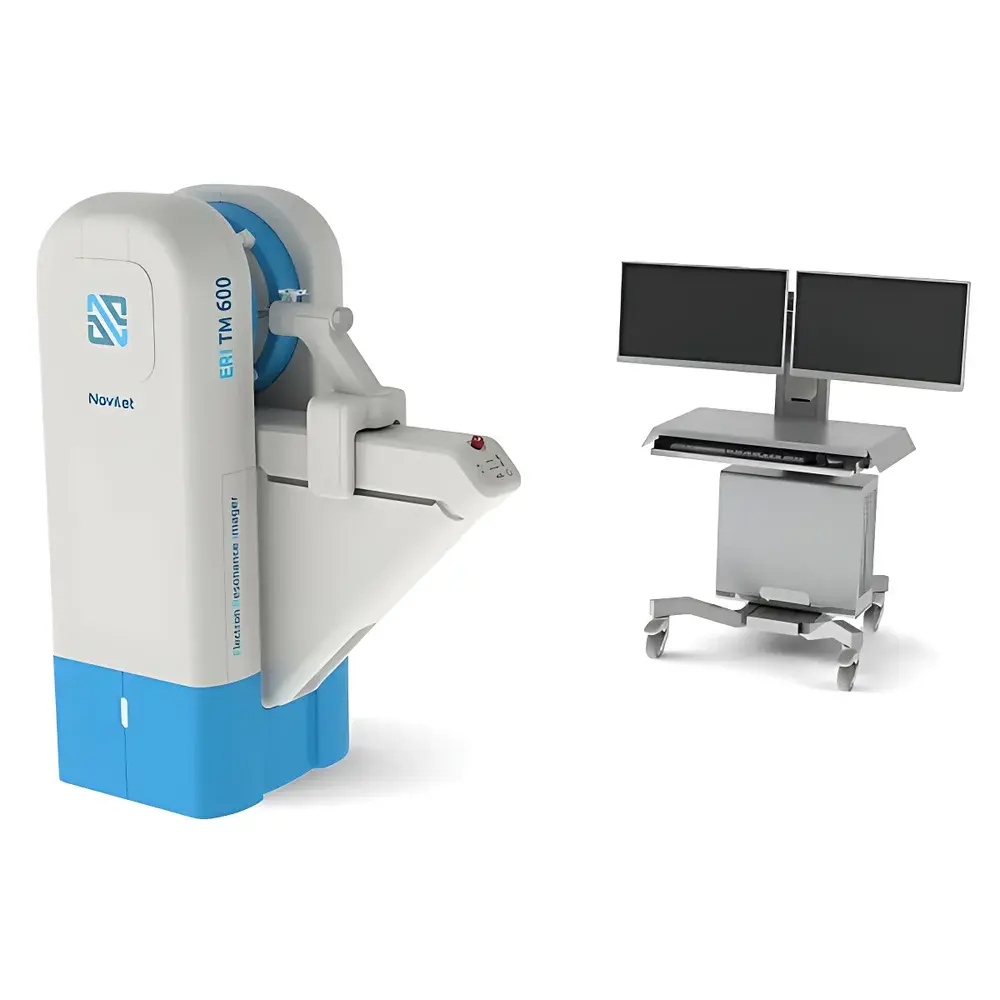
| Brand | Novilet |
|---|---|
| Origin | Poland |
| Model | ERI TM 600 |
| Imaging Modality | Electron Paramagnetic Resonance (EPR) Imaging |
| Sample Type | Live small animals (mouse, rat) |
| Spin Probe Required | Yes |
| Field Strength Range | 0.01–500 G (CW), 0.01–50 G (rapid scan) |
| Operating Frequency | 575 MHz |
| RF Power Output | Up to 500 mW |
| Modulation Amplitude | Up to 50 G |
| Modulation Frequency | 1 kHz |
| Magnetic Field Resolution | 10 mG |
| Field Stability | ±20 mG |
| Field Noise | ≤5 mG RMS |
| Gradient Strength | 13 G/cm |
| Magnet Bore Diameter | 34 mm |
| Imaging Volume | 20 cm³ |
| 3D Acquisition Time | As low as 4.5 s (225 projections) |
| SNR (OXO63, 25 g mouse, 100 ms) | 112 |
| Digital Resolution | 16-bit |
| Q-Factor | >800 |
| Temperature-Controlled Animal Holder | Integrated |
| Anesthesia-Compatible Gas Exchange System | Included |
| Stereotactic Mouse Brain Fixation | Standard |
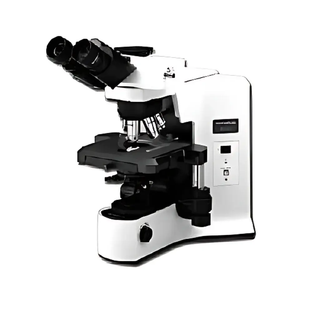
| Brand | OLYMPUS |
|---|---|
| Origin | Japan |
| Model | BX41 |
| Light Source | 6 V / 30 W Halogen Illuminator |
| Optical System | UIS2 (Universal Infinity System) |
| Observation Tube Options | Binocular, Tilting Binocular, Trinocular |
| Objective Turret | 5-Position Revolving Nosepiece |
| Compliance | ISO 9001, JIS B 7151, CE Marked for In Vitro Diagnostic Use |
| Software Compatibility | Supports Olympus cellSens™ and third-party DICOM/GLP-compliant image acquisition platforms |
| Regulatory Status | Designed for research use only (RUO), not for clinical diagnostic use per FDA 21 CFR §809.3(a) |
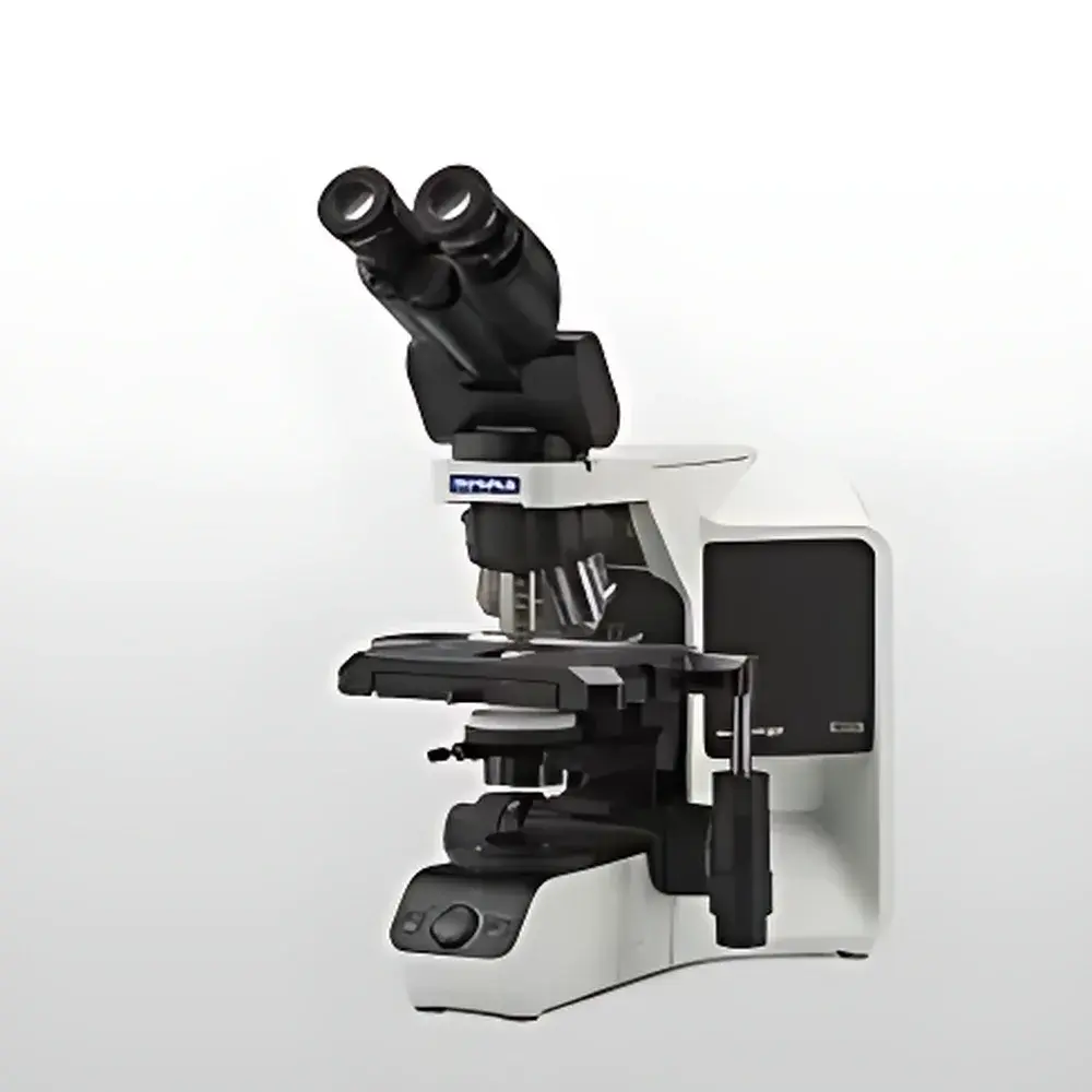
| Brand | Olympus |
|---|---|
| Origin | Japan |
| Manufacturer | Olympus Corporation |
| Product Type | Upright Microscope |
| Model | BX43 |
| Light Source | High-CRI White LED (equivalent to 30 W halogen, stable color temperature across intensity levels, 20,000 h lifetime) |
| Ergonomic Design | Adjustable observation tube and mechanical stage |
| Modularity | Fully modular optical platform for configuration scalability |
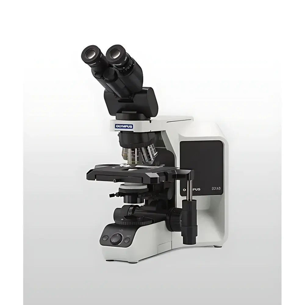
| Brand | Olympus |
|---|---|
| Origin | Japan |
| Model | BX43 |
| Type | Upright Biological Microscope |
| Illumination | LED Transmitted Köhler Illuminator (White Light, 6200 K, 20,000 hr lifetime) + 100 W Mercury Arc Lamp for Fluorescence (UV/Blue/Green Excitation) |
| Observation Modes | Brightfield, Darkfield, Simple Polarization, Differential Interference Contrast (DIC), Fluorescence |
| Objective Turret | Manual or Motorized 7-Position |
| Stage | Mechanical Right-Hand Control, 76 × 52 mm Travel (X/Y) |
| Condenser Options | Abbe (NA 1.1), Achromat/Apochromat (NA 1.4, Oil Immersion), Swing-out Darkfield (Dry/Oil), Polarizing, Phase Contrast-Compatible |
| Eyepiece Field Number | FN 22 or FN 26.4 |
| Dimensions (W×D×H) | 274.5 × 362 × 410 mm |
| Weight | 13 kg |
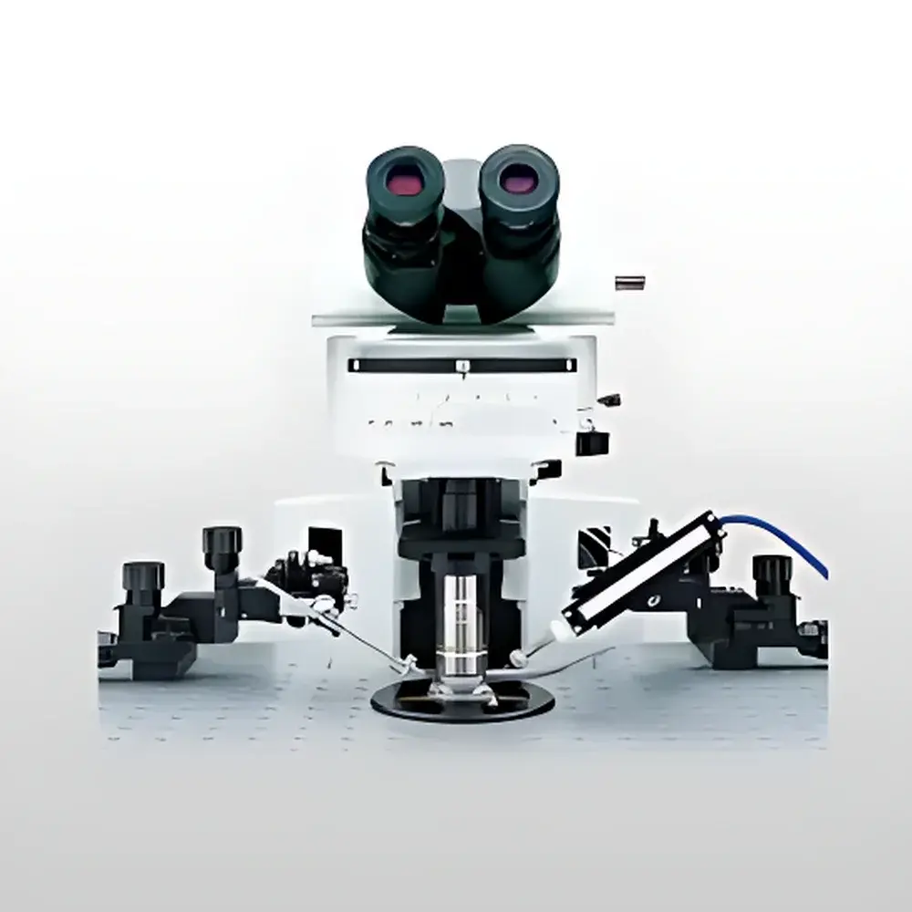
| Brand | OLYMPUS |
|---|---|
| Origin | Japan |
| Manufacturer | OLYMPUS Corporation |
| Product Type | Upright Fluorescence Microscope |
| Excitation Source | 100 W Mercury Arc Lamp |
| Illumination | Fiber-Optic Light Guide |
| Observation Head | Trinocular |
| Focus Mechanism | Manual Coarse/Fine Adjustment |
| Stage Type | Fixed Mechanical Stage |
| DIC Capability | Nomarski Differential Interference Contrast (DIC) Enabled |
| Fluorescence Filter Turret | 6-Position Motorized or Manual Turret with Click-Stop Release |
| Objective Mount | WI-SRE3 Swing-Out, U-SLRE Slide-Out, or WI-SNPXLU2 Single-Objective Nosepiece |
| Objective Compatibility | High-NA Water-Immersion (e.g., XLUMPLFLN20xW), XLFLUOR Series (2x/340, 4x/340), and Standard RMS-thread Objectives |
| Condenser Options | Multiple DIC-Compatible Condensers (e.g., U-UCD8) |
| Vibration-Damping Frame | Integrated Anti-Vibration Optical Bench Architecture |
| IR Transmission | Optimized for Near-Infrared (NIR) Imaging up to ~900 nm |
| Compliance | Designed for GLP/GMP-aligned electrophysiology and in vivo imaging workflows |
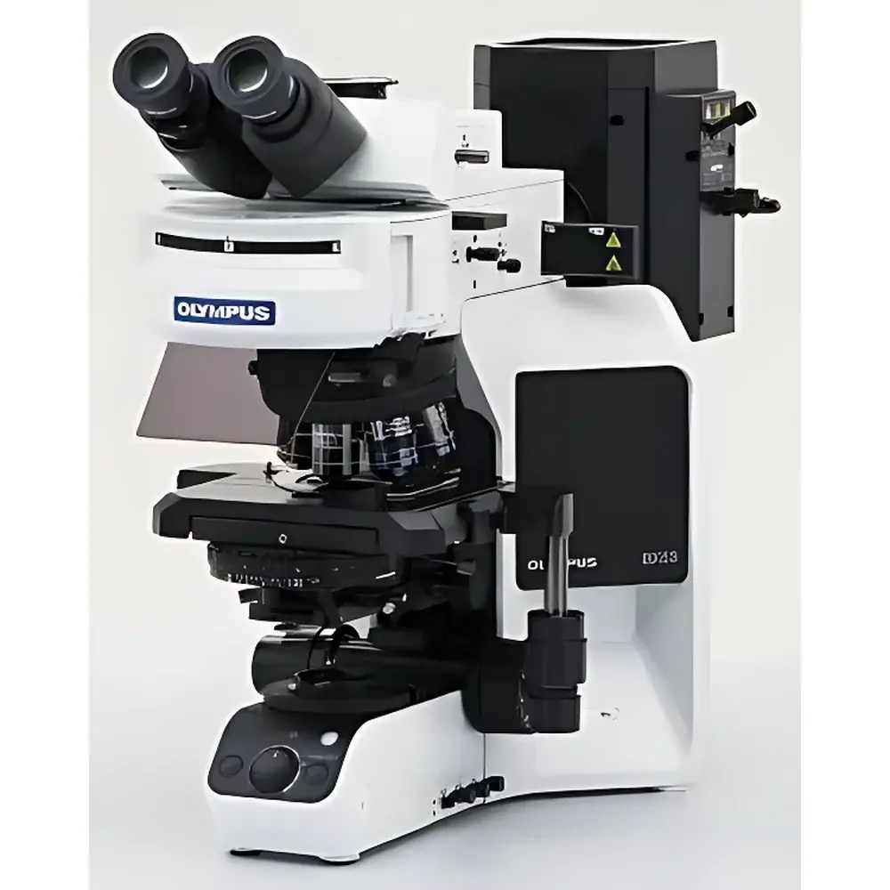
| Brand | Olympus |
|---|---|
| Origin | Japan |
| Instrument Type | Upright Biological Microscope |
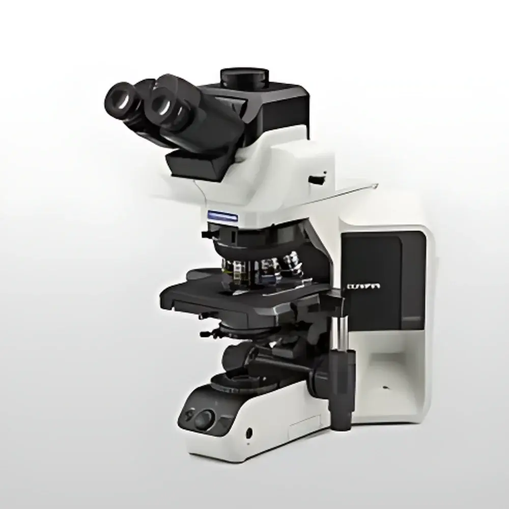
| Model | BX53 |
| Illumination | 100 W Mercury Arc Lamp (Fluorescence), Halogen/LED (Transmitted Light) |
| Objective Turret | Manual or Motorized 7-Position |
| Stage | Mechanical Right-Hand Control, 76 × 52 mm Travel |
| Condenser | Motorized Universal Condenser (NA 0.9–1.4, WD 0.63–2.0 mm) |
| Observation Modes | Brightfield, Darkfield, Phase Contrast, Simple Polarization, DIC, Fluorescence (UV, Blue, Green Excitation) |
| Eyepiece Field Number | FN 22 or FN 26.5 |
| Dimensions (W×D×H) | 274.5 × 614 × 469 mm |
| Weight | 21 kg (Fluorescence Configuration) |
| Brand | OLYMPUS |
|---|---|
| Origin | Japan |
| Manufacturer Type | Original Equipment Manufacturer (OEM) |
| Origin Category | Imported |
| Model | BX53(LED) |
| Instrument Type | Upright Fluorescence Microscope |
| Excitation Source | High-Intensity LED |
| Instrument Class | Conventional Fluorescence Microscope |
| Objective Options | Multiple X Line Objectives (e.g., 4×, 10×, 20×, 40×, 60×, 100× oil) |
| Fluorescence Modules | Interchangeable Filter Cube Turrets (e.g., FITC, TRITC, DAPI, Cy5) |
| Illumination | 100 W Mercury Arc Lamp (optional), Fiber-Optic Light Guide (optional), Integrated LED Illuminator (standard) |
| Control Mode | Manual and Motorized |
| Focusing Mechanism | Coaxial Fine/Coarse Focus with Motorized Z-Drive Option |
| Condenser Options | U-UCD8, U-LC, U-ULC, U-SC3 |
| Observation Heads | Binocular or Trinocular (with C-mount port) |
| DIC Components | Available as Add-on Module (U-DICR, U-DICB) |
| Stage | Mechanical XY Stage (76 × 52 mm travel), Optional Motorized Stage |
| Fluorescence Filter Sets | Broadband and Narrowband Bandpass Filters, Dichroic Mirrors, Emission Filters (all standardized to ISO 10934-1) |
| Brand | Olympus |
|---|---|
| Origin | Japan |
| Manufacturer Type | Original Equipment Manufacturer (OEM) |
| Origin Category | Imported Instrument |
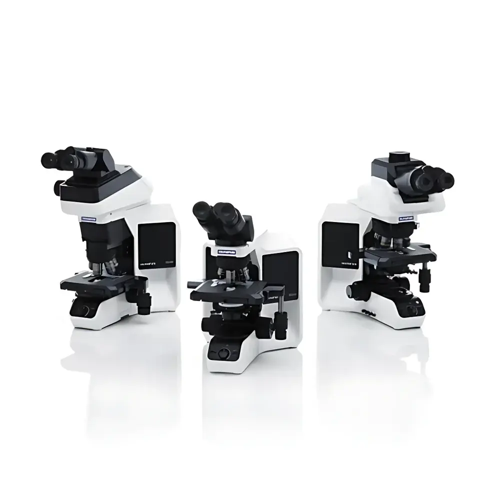
| Model Series | BX53(LED), BX43, BX46 |
| Instrument Type | Upright Microscope |
| Illumination Source | High-CRI White LED |
| Design Focus | Ergonomic Clinical Workflow Optimization |
| Brand | Olympus |
|---|---|
| Origin | Japan |
| Manufacturer Type | Original Equipment Manufacturer (OEM) |
| Origin Category | Imported Instrument |
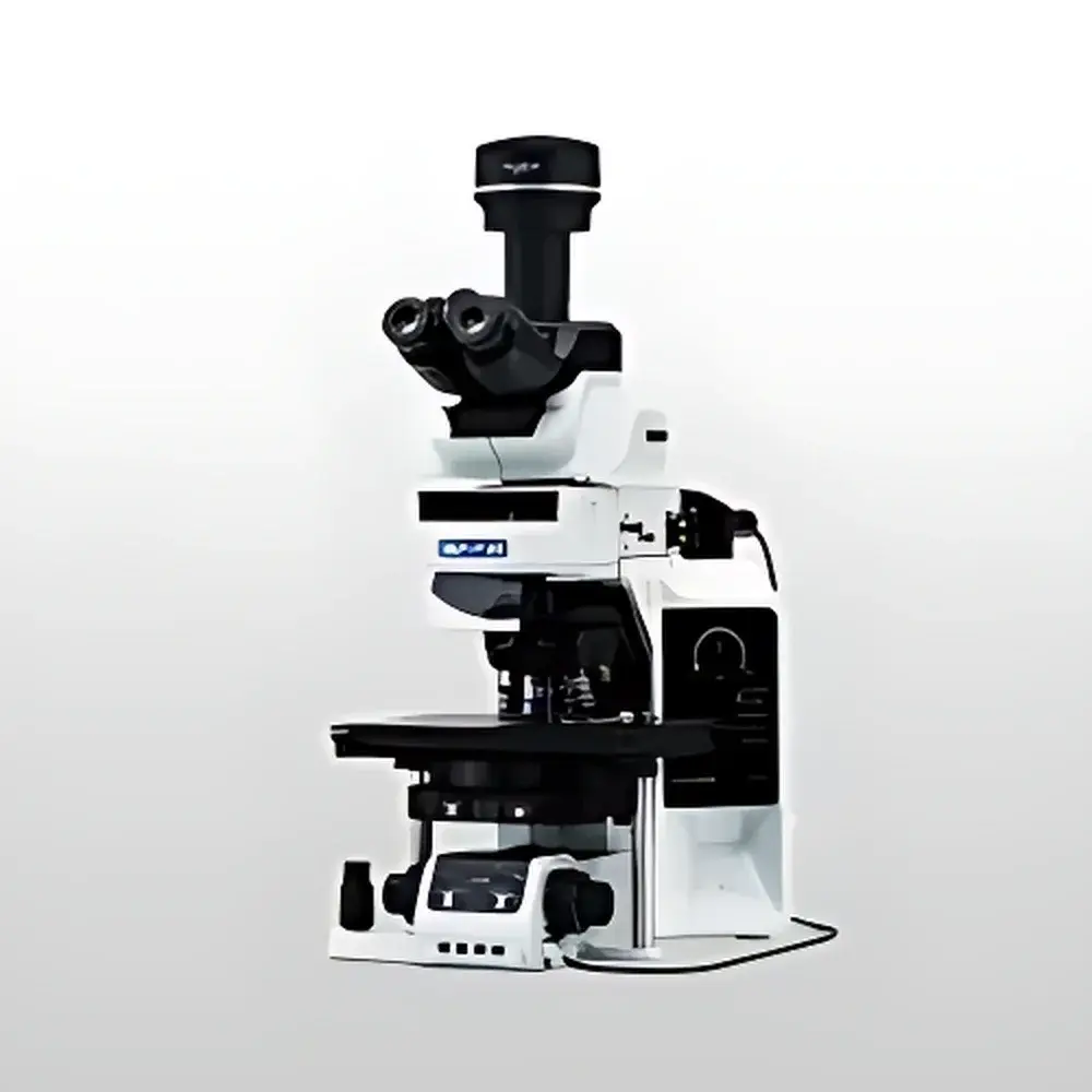
| Model | BX63 |
| Instrument Type | Upright Fluorescence Microscope |
| Instrument Class | Research-Grade Fluorescence Microscope |
| Objective Turret | Motorized |
| Focus Mechanism | Manual and Motorized |
| Condenser Control | Manual and Motorized |
| Observation Head | Binocular and Trinocular |
| DIC Compatibility | Integrated Differential Interference Contrast (DIC) Module Available |
| Stage | Manual, Mechanical, and Ultrasonic Piezo Motorized XY Stage |
| Fluorescence Filter Sets | Multiple Standard and Customizable Excitation/Emission Filter Combinations |
| Control Interface | Touchscreen Controller with Preset Recall and Workflow Manager |
| Brand | Olympus |
|---|---|
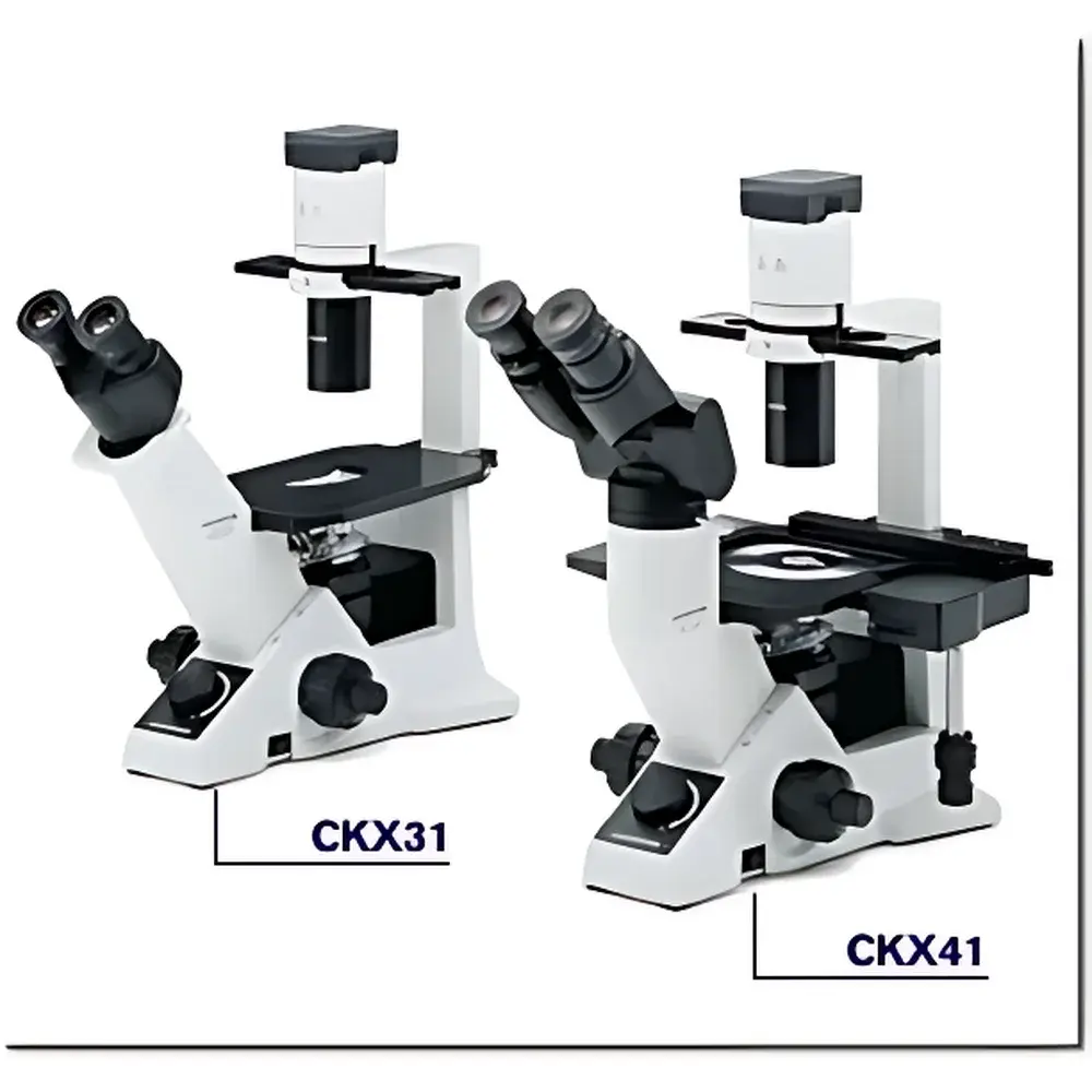
| Model | CKX31 |
| Optical System | UIS2 Infinity-Corrected |
| Illumination | 6V/30W Halogen Lamp with Heat-Absorbing Filter and Ground Glass Diffuser |
| Objective Turret | 4-Position Revolving Nosepiece |
| Stage | Right-Hand Low-Position Coaxial X-Y Mechanical Stage (X=120 mm, Y=78 mm) |
| Observation Tube | 30° Inclined Binocular Tube (IPD: 48–75 mm) |
| Objectives | 4× Plan Achromat, 10× Phase Contrast (PH1), 20× Long Working Distance Phase Contrast (PH1) |
| Eyepieces | 10× Widefield (FN 20) |
| Optional Accessories | Mechanical Stage Extension, Fluorescence Illuminator, CCD Imaging System |
| Compliance | Designed for ISO 9001-certified manufacturing environments |
| Brand | OLYMPUS |
|---|---|
| Origin | Japan |
| Model | CKX41, CKX53 |
| Microscope Type | Inverted Microscope |
| Eyepiece Configuration | Trinocular |
| Optical System | UIS2 Infinity-Corrected Optics |
| Focusing Mechanism | Objective Nosepiece-Driven Coaxial Coarse/Fine Focus (Fixed Stage) |
| Total Focus Travel | +7 mm / –2 mm from 1 mm above stage surface |
| Coarse Focus Travel per Revolution | 39.6 mm |
| Fine Focus Travel per Revolution | 0.2 mm |
| Fine Focus Resolution | 2 µm |
| Objective Turret | 4-Position |
| Stage | Mechanical XY Stage (X = 120 mm, Y = 78 mm) with Removable Circular Insert (Ø25 mm) and Petri Dish Support (Ø35 mm) |
| Condenser | Removable Long Working Distance Condenser (NA 0.3, WD 72 mm) |
| Phase Contrast | Pre-centered and Centerable Phase Contrast Slides (PHC-Compatible Objectives: CPLN10×PH, CPLFLN10×PH, LCACHN20×PH) |
| Fluorescence Illumination | Vertical Fluorescence Unit with 50 W Mercury Lamp, Three-Position Filter Slider (B, G, Transmitted Light or U), Optional U-Filter Block |
| Eyepiece Options | Widefield WH10× (F.N. 22), WHB10× (F.N. 20), PWH10×, 35WH10× |
| Tube Options | U-CTR30-2 Trinocular Tube (30° inclination, 50–75 mm IPD, beam splitter 50/50 or 20/80), CKX-TBI Adjustable-Incline Binocular Tube (30°–60°), UTBI-3 (5°–35°) |
| Power Supply | Dual Voltage Auto-Switching (100–240 V AC, 50/60 Hz), Continuously Variable Intensity Control |
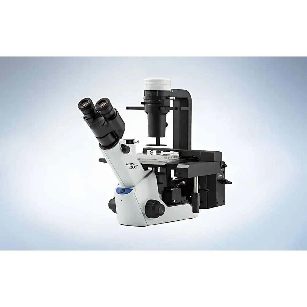
| Brand | Olympus |
|---|---|
| Origin | Beijing, China |
| Model | CKX53 |
| Instrument Type | Inverted Microscope |
| Optical System | UIS2 |
| Illumination | Long-life LED (white light), Optional 100W Mercury Lamp (U-LH100HG) for Fluorescence |
| Phase Contrast | Integrated iPC (intelligent Phase Contrast) System with Universal Annulus Plate for 10×, 20×, and 40× Objectives |
| IVC (Inverse Contrast) Technology | Enabled with PLCN10× or CACHN10×IPC Objective |
| Field Number | FN 22 |
| Weight | ~7 kg |
| Stage Compatibility | Standard CKX-MVR Manual Stage |
| Fluorescence Mirror Units | Interchangeable, Compatible with IX3/BX3 Series Filter Sets |
| UV-Resistant Coating | Yes |
| Sterility-Friendly Design | Bottom-mounted shutter, compact footprint, cleanbench-compatible |